whiplash
Fig. 1 Whiplash – Mechanism of Injury

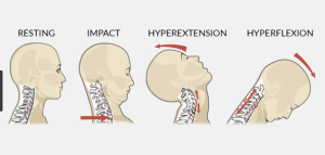
Acceleration/deceleration injury to the cervical spine(neck) and back, also known as whiplash, is a common injury due to traffic accidents regularly treated by our physiotherapists in Tralee. Whiplash as it is commonly known may result from  rear-end or side impact motor collisions. It can also occur with activities such as diving or due to a direct blow from an opponent etc. The impact of any of these may result in injury to the cervical spine(bones of neck) or soft tissues such as the muscles or ligaments of the neck/back. This in turn leads to what is called ”whiplash associated disorders” or WAD for short(Spitzer, W.O., 1995).

To simplify the mechanism of this type of injury, think of a whip snapping. This is basically what is happening with your neck during these incidents, the head jolts swiftly forward first, then snaps backwards. It is being propelled swiftly with great force in one direction, then there is a recoil swiftly in the opposite direction. The back may also experience similar trauma.

The most common symptoms of whiplash include neck pain, headache, back and shoulder pain with decreased mobility. The condition can be graded into WAD 1 – 4 depending on the degree on injury. With Wad 1 the patient complaints of pain, stiffness or tenderness only but no physical signs are noted. With Wad 2 the patient complains of neck pain and there is decreased range of motion and point tenderness in the neck. Where there are hard neurological signs, for example tingling and/or numbness down one or both arms WAD 3 is the grade assigned. Where there is a resulting fracture of the cervical spine, the term WAD 4 is used(Balla and Iansek, 1988,  Stovner, L.J., 1996).

With whiplash the patient may not feel any pain immediately after the accident, however symptoms can increase gradually in the 48 hours after  injury. Muscles, joints, ligaments and neural tissue can all be affected depending on the level of injury. Once an x-ray or Mri is performed to rule out serious injury to the bones, ligaments etc. of the neck , red flag signs also being ruled out, it is important that physical  therapy is started as soon as possible. A multimodality approach to treatment as early as possible has shown good results in best evidence practice. This includes very specific therapeutic exercises, manual therapy, postural education and the use of drugs most notably non steroidal anti-inflammatories and muscle relaxants(Childs et al. 2008, 2009). It is extremely important to try to return range of movement to the neck as soon as possible after the accident once adequate safety checks have been performed. The neck stiffens up quite quickly when immobilized for even short periods of time. This current thinking is very different to years ago when neck braces and immobilization for long periods after accidents was standard.

Some symptoms  associated with whiplash that can indicate the condition may be a little more serious, needing immediate review by a doctor or specialist include bilateral parasthesia(numbness on both sides of body), dizziness, feeling sick, a lump in throat when swollowing, progressively worsening neurological symptoms, signs of neck instability, unrelenting pain, increase in any of these symptoms with rotation or flexion of head. For more information on whiplash click here.


References

Balla J., Iansek, R., Headaches arising from disorders of the cervical spine. In : Hopkins A, ed. Headache. Problems in diagnosis and management. London: Saunders, 1988:241-67.

Childs, J.D., Cleland, J.A., Elliott, J.M. et al. Neck pain: clinical practice guidelines linked to the International Classification of Functioning, Disability, and Health from the Orthopedic Section of the American Physical Therapy Association. Erratum appears in J. Orthop. Sports Phys. Ther. 2009 Apr;39(4):297, J. Orthop. Sports Phys. Ther. 2008;38(9):A1-34.

Spitzer, W.O., Skovron, M.L., Salami, L.R. et al. Scientific monograph of the Quebec Task Force on Whiplash Associated Disorders : redefining ”whiplash” and its management. Spine 1995;22(8suppl.):S1-73.

Stovner, L.J. The nosologic status of the whiplash syndrome: a critical review based on a methodological approach. Spine 1996; 21(23):2735-46.


thoracic spine mobility
Thoracic Spine

A mobile thoracic spine allows you turn your back in many directions, enabling you to do everyday tasks with ease. Today’s sedentary lifestyle often contributes to reduced spinal mobility. Basically ”motion is lotion” and if you are inactive and also prone to poor posture, your thoracic spine can seize up. If it goes on long enough, say into old age portions of the spine may fuse and not move at all. A lack of thoracic spine mobility means that the lumbar spine, pelvis, shoulders and surrounding muscles  have to compensate. Long term, these over-compensations can  lead to overuse conditions and injuries, the lower back being particularly suceptable. This is because the lumbar spine is meant to keep us stable and is not very mobile, so when these joints are forced to overcompensate for the lack of movement in the thoracic area, it can place alot of pressure on the discs of the lower back. Possible consequences include inflammation, degeneration,  herniation of the discs, generalized low back pain, compression fractures, muscles spasms, and spinal nerve injuries. Similar pressures and injuries can occur in the neck and shoulders. For example, if your thoracic spine isn’t mobile, anytime you have to do a movement overhead, your shoulders make up for that lack of mobility. If you have shoulder impingement or chronic shoulder and neck problems lack of mobility in the thoracic spine will make every thing worse.


Improving Thoracic Spine Mobility

Yoga, pre- and post-workout stretching, and mobility exercises are the best way of maintaining and improving thoracic spine mobility. These need to be done regularly and consistently, especially as you get older. Your physiotherapist will be able to guide you on the correct exercises and help correct your form and technique until you get used to them and can do them yourself. Here are a few exercises to get you started.

For more information contact us @ Physiotherapists in Tralee.


Chondromalacia Patella – Knee Pain – Patellar Tracking Dysfunction

Chondromalacia patella
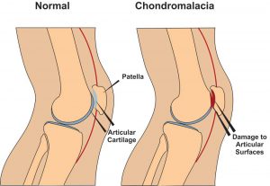
Chondromalacia patella, also known as ”Runners Knee” is one of the most common causes of knee pain in runners. The condition results from irritation of the cartilage on the under-surface of the kneecap. This cartilage is smooth and the kneecap normally glides effortlessly across it during bending of the knee joint. In some individuals however the kneecap does not track so smoothly due to poor alignment and the cartilage surface becomes irritated, resulting in inflammation and knee pain. In more severe cases there can be breakdown of the cartilage. Chondromalacia patella can affect athletes of any age but tends to be more common in women, most likely due to anatomical differences between the sexes ie. wider hips in females which results in a greater angulation between hip and knee, thus resulting in increased lateral forces on the patella.


Chondromalacia Patella – Causes

There are several causes both structural and dynamic which are linked to the condition. These include excessive foot pronation(feet turn out when running etc.), tight IT band, tight vastus lateralis(basically outer lower quad), weak or slow firing vastus medialis (basically lower inner quad), increased Q angle (simply put the angle between the outer hip and centre of the knee), a lateral femoral condyle that is not sufficiently prominent anteriorly (simply put the knee joint does not fit together properly),and a small or high riding patella(knee cap).(McConnell, 2002)


Chondromalacia Patella – Symptoms

The most common symptom is a dull, aching pain in the front of the knee, behind the kneecap. This pain is often worse when you go up or down stairs. It also can flare up after you have been sitting in one position for a long time. For example, your knee may be painful and stiff when you stand up after watching a movie or after a long trip in a car or plane. In some cases, the painful knee also can appear puffy or swollen. Chondromalacia can sometimes cause a creaky sound or grinding sensation known as ”crepitus” when you move your knee.


Chondromalacia Patella – Physio Treatment 

Suitable treatment may involve 1. Soft tissue work to loosen tightened structures such as vastus lateralis muscle, IT band, lateral retinaculum etc., 2.Strengthening of weak structures such as vastus medialis, glutes , hip abductors etc., 3. Correction of overpronation using orthotics, 4. Non steroidal anti-inflammatories such as ibuprofen to reduce pain and inflammation, 5. Rest with gradual return to exercise, 6. Taping to correct tracking can be a short term solution.(Hertling and Kessler, 2006) while you strengthen the vastus medialis muscle. Also there are supports you can purchase to help correct patella tracking while exercising. These are a good short term solution while you correct the problems referred to above. Here is a good example.

If nonsurgical treatments fail, or if you have severe symptoms, your doctor may recommend arthroscopy to check the cartilage inside your knee. If the cartilage is softened or shredded, damaged layers can be removed during the surgery, leaving healthy cartilage in place .


References

Hertling, D., Kessler, R.M. ”Management of Common Musculoskeletal Disorders : Physical Therapy Principles and Methods.” Lippincott, Philidelphia 524-533, 2006.

Mc Connell, J. ”The physical therapist’s approach to patellofemoral disorders.” Clinical Sports Medicine 21:363-387, 2002.


For more information on chondromalacia patella see this video.


If  you would like to get in touch to discuss your condition or make an appointment for physio in Tralee, please click

here for details.

Regards Eddie.

Back Mice
Fig. 1 Back Mice

The term “back mice” is a rather cute description for a painful yet often overlooked condition, even by back pain specialists. The term  “back mice” was first used to label the condition by Peter Curtis in 1993. Back mice present as small, firm, fleshy yet moveable nodules upon palpation over the sacral region. Firm pressure directly on the nodules usually produces pain and tenderness, which sometimes radiates into the sacrum and hip. Also, the back mouse seems to suddenly appear following trauma to the back as in a motor vehicle accident or perhaps following a lifting injury. The size of the nodules does not change and they remain the same regardless of the administered soft tissue treatment.

Perhaps a more descriptive term for the back mouse is  “lumbar fascial fat herniation” . This occurs when the lumbar subfascial fat layer herniates through the overlying thoraco-dorsal fascia and gets trapped and inflamed. The mechanism appears to be due to an anatomical defect or weakened area in the fascia, which, when there is increased internal pressure, allows the fat lobules to push through the fascia. Once herniated, the fat becomes trapped and as an expanded, inflamed, herniation in an otherwise unyielding fibrous capsule. This creates a focus of pain. Pressure on the fat mouse does not push it back through the fascia but only inflames the torn fascia more. These herniations occur at predictable sites along the iliac crest and sacrum very close to the natural dimple area. They also are approximately three times more prevalent in women, particularly in moderately obese women.

A client usually presents with an episode of  low back pain. There may be a history of pain with lifting or prolonged sitting and the pain is usually greater on one side more than the other. The pain may radiate into the buttocks and sacrum and perhaps to the lateral thigh and into the lower extremity. Medications usually do nothing. Many will have gone through the pain pill merry-go-round, taking a cocktail of pain and anti-inflammatory medications even though the pain never truly goes away. The client has often tried everything, been everywhere and you are their last hope. They may have had radiographs, MRI studies or nerve conduction studies, all with negative or minimal findings. They may even have a minor disc bulge without nerve compression, yet the pain exactly mimics a discogenic disorder. When asked to point to the area of the greatest pain, they will invariably point just above and lateral to the natural “dimple” where the back and buttocks come together, near the multifidis triangle. When the area is palpated, the most marked finding is one or several firm, mobile 1.3 cm nodules. When pressed, these nodules reproduce the client’s complaints of back pain as well as the “sciatic” pain.

Subcutaneous lipomas in the back region differ from back mice in that they present as moveable non-tender “speed bumps” that cause pain only when they compress the underlying soft tissue. Subcutaneous lipomas can be found anywhere in the body. They grow slowly over time and are only cosmetically important.

back mice
Fig 2. Overlying thoraco-dorsal fascia.


back mice
Fig 3. Iliac crest and sacrum.


There always seems to be a focus by back pain specialists upon the disc and nerves issues even though the fat mice are readily palpable. Many sufferer’s have had epidural injections without success. I have had clients who have had surgery for disc herniations yet who still point to the back mouse post surgery as the focal point of pain. It could perhaps be considered diagnostic for the presence of a lumbar fascial fat herniation if a local infiltration of anesthetic takes away the pain.

Treatment of Back Mice

A medical doctor can inject the back mouse with a local anesthetic. This usually only helps temporarily. Dry needling techniques by acupuncturists may help reduce the tension in the fibrous capsule. Good results may sometimes be obtained with local electrical stimulation techniques such as electro-acupuncture. Do not apply deep pressure to back mice during manual physiotherapy treatment. Doing so may only serve to aggravate the herniation. Release of muscular tissue tension around the back mouse may provide some relief by easing pressure on the area.

Perhaps the only permanent cure for the back mouse is its excision and removal. This could be performed by a hernia repair specialist. Once the fat herniation is excised and the fascial tear repaired, the client usually enjoys a more enduring and sometimes dramatic relief. One of the biggest problems is that so many medical doctors fail to recognize this condition; they tend to discount its existence, thereby limiting the treatment options. Icing the area may also provide temporary relief.


For a short video on back mice click here.


Physiotherapist in Tralee. Phone 0867700191 to discuss your injury, make an appointment or get a second opinion.


Achilles tendon ruptures will debilitate your lower leg completely, the moment they happen. The Achilles tendon is an important part of the leg. It is located just behind and above the heel, attaching the heel bone to the calf muscles. Its function is to help in bending the foot downwards at the ankle (this movement is called plantar flexion). If the Achilles tendon is torn/ruptured, the tear may be either partial or complete. In a partial tear, the tendon is torn but is still partly joined to the calf muscle. With complete tears, the tendon is completely torn and the connection between the calf muscles and the ankle bone is lost. If your Achilles tendon is ruptured you will be unable to stand on your tiptoes and you will have a flat-footed walk with a severe limp, along with considerable pain initially.


An Achilles tendon can tear when there is too high a load or stress placed on it. This can happen with activities involving a forceful push off with the foot – ie. running/sprinting in football, basketball, tennis etc. I have also seen it occur when somebody sprints off suddenly on a sandy beach, the sand giving way under the foot putting extra stress on the calf and the achilles tendon. The push off movement uses a strong contraction of the calf muscles maximally stressing both the calf muscles and the Achilles tendon . Injury to the achilles can also occur due to falls, if the foot is suddenly forced into an upward-pointing position, stretching the tendon. Another possible injury is a deep cut/laceration to the tendon.


Achilles Tendon Rupture
   Fig. 1.  Achilles Tendon Rupture


Sometimes the Achilles tendon is weak, making it more prone to rupture. Factors that weaken the Achilles tendon are:

  • Corticosteroid medications (such as prednisolone) – mainly when used as long-term treatment.
  • A corticosteroid injection near the Achilles tendon.
  • Certain rare medical conditions, such as Cushing’s syndrome, where the body makes too much of its own corticosteroid hormones.
  • Tendinopathies of the Achilles tendon.
  • Other medical conditions which can make the tendon more prone to rupture; for example, rheumatoid arthritis, diabetes, gout and systemic lupus erythematosus.
  • Certain antibiotic medicines may slightly increase the risk of having an Achilles tendon rupture. These are the quinolone antibiotics such as ciprofloxacin and ofloacin. The risk of having an Achilles tendon rupture with these antibiotics is quite low and mainly applies to people who are also taking corticosteroid medication.


You might hear a snap or feel a sudden sharp pain when the tendon is torn (ruptured)during a sporting activity or injury. The sharp pain usually settles quickly, although there may be some aching at the back of the lower leg. After the injury, the usual symptoms are:

  • A flat-footed type of walk. You can walk and bear weight but cannot push off the ground properly on the side where the tendon is ruptured.
  • Inability to stand on tiptoe.
  • If the tendon is completely torn, you may feel a gap just above the back of the heel. However, if there is bruising and swelling may disguise the gap.

An Achilles tendon rupture is usually diagnosed based on symptoms, history of the injury and a physio/doctor’s examination. An orthopedic test called Thompson’s test (also known as the calf squeeze test) may help diagnosis. In this test, you will be asked to lie face down on the examination bench and to bend your knee. The doctor will gently squeeze the calf muscles at the back of your leg and observe how the ankle moves. If the Achilles tendon is OK, the calf squeeze will make the foot point away from the leg (a movement called plantar flexion). This is quite an accurate test for Achilles tendon ruptures. An ultrasound or MRI may be used to confirm diagnosis and give a fuller picture of the injury.


Treatment and recovery

Treatment for a ruptured achilles tendon usually involves surgery to repair the tendon. The surgeon sews/sticks together the torn ends of the Achilles tendon, and may also use another tendon or a tendon graft to help with the repair. A plaster cast or brace is needed after the operation for about eight weeks, to keep the foot immobilized, allowing the tendon can heal. The plaster cast or the brace is positioned so that the foot is pointing slightly downwards, which takes the strain off the tendon.

Traditionally, crutches were used to keep weight off the leg during the first few weeks of treatment. Current thinking tends towards using the leg normally early on (early mobilization). This involves fitting a plaster cast or a brace which you can walk on. It is more convenient because you do not need to use crutches. Physiotherapy will also be needed, especially when the cast is removed. I have personally found instrument assisted soft tissue work(after about 8 weeks) to be very beneficial along with stretching/strengthening work and deep tissue work to the calf during recovery. Full recovery is greatly slowed when there is no hands-on work done during recovery, after the cast is removed once an ok is given by the surgeon to commence.

Depending on a person’s profession, some people may need several weeks off work after an Achilles tendon tear (rupture); the time taken to return to sport is usually between 4 and 12 months. Generally, the outlook is good. However, the tendon does take time to heal, usually about six to eight weeks. More time will be needed after this to allow the muscles and tendon to regain normal strength.


For more on rehabilitation on achilles tendon ruptures post surgery click here.


Physiotherapist in Tralee. Ring 08677001 to discuss your condition, book an appointment or get a second opinion.

Herniated Disc vs Bulging Disc

herniated disc
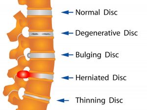
Your spine is made up of lots of bones called vertebrae and discs stacked on top of one another, forming the spinal canal. The nerves of the spinal cord run down the length of the spinal canal. The discs in act like shock absorbers between the vertebrae. These discs are made of two components: a softer center (called nucleus pulposus) surrounded by a tough elastic-like band (called annulus fibrosus). A bulging disc is like a squashed jam doughnut before the jam bursts out. The disc sags and looks like it is bulging outward. With a herniated disc, the outer covering of the disc has a hole or tear. This causes the nucleus pulposus (jelly-like center of the disc) to leak into the spinal canal. It is like the jam leaking out from the inside of a squashed jam doughnut.
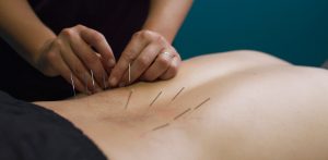
Bulging discs are usually caused by age-related degeneration. There is usually a progressive, gradual onset of symptoms. Bulging discs and herniated discs can occur anywhere along the spine. Pain from a herniated disc usually comes on abruptly often affecting one individual nerve root. Herniated discs are often caused by an acute injury. In some cases, you may know the cause of the injury, such as twisting incorrectly.

Herniated and bulging discs are the most common causes of lower back pain, as well as leg pain or ”sciatica.” Between 60% and 80% of people will experience low back pain at some point their lives. Although a herniated disk can be very painful, most people feel much better with just a few weeks or months of nonsurgical treatment, such as using  muscle relaxants and anti-inflammatory medications, along with physiotherapy.  Long term management of these conditions needs to including properly prescribed strengthening and flexibility exercises in order to avoid surgery. Surgery is the final option.

For more on herniated and bulging discs, see video.


Physio in Tralee. Phone 0867700191 to discuss your condition or make an appointment.

Dry-NeedlingDry Needling

Trigger-point dry needling is a procedure where a fine  acupuncture needle is inserted into the skin and muscle. It is aimed at myofascial trigger points, which are points of exquisite pain in skeletal muscle, that are associated with a hypersensitive palpable nodule or a taut band.

Active trigger points can spontaneously trigger local or referred pain. They cause muscle weakness, restricted range of movement and autonomic phenomena. Latent trigger points do not cause pain unless they are stimulated. They may alter muscle activation patterns and contribute to restricted range of movement . Therefore both active and latent trigger points cause allodynia(nerve pain) at the trigger point site and hyperalgesia away from the trigger point following applied pressure.

The formation of trigger points is caused by the creation of a taut band or knot within the muscle. This band is caused by excessive acetylcholine release from the motor endplate combined with inhibition of acetylcholine esterase and upregulation of nicotinic acetylcholine receptors.  Motor end plates, also called neuromuscular junctions, are specialised chemical synapses formed at the sites where the terminal branches of the axon of a motor neuron contact a target muscle cell. Motor neurons are nerve cells that send electrical output signals to the muscles.

Initially the taut bands are produced as a normal protective, physiological measure in the presence of actual or potential muscle damage. They are thought to occur in response to unaccustomed eccentric or concentric loading, sustained postures and repetitive low load stress. However when sustained they contribute to sustained pain. The pain caused by trigger points is due to hypoxia and decreased blood flow within the trigger point. This leads to a decreased pH which activates the muscle nociceptors to restore homeostasis. This causes peripheral sensitization. Trigger points are also involved in central sensitization. The mechanism remains unclear but trigger points maintain nocioceptive input into the dorsal horn and therefore contribute to central sensitization.


Stimulation of a local twitch response (LTR)

Dry-needling of these myofascial trigger points via mechanical stimulation causes an analgesic effect. This mechanical stimulation causes a local twitch response (LTR). A LTR is an involuntary spinal cord reflex contraction of the muscle fibers in a taut band. Triggering an LTR has been shown to reduce the concentration of nociceptive substances in the chemical environment near myofascial trigger points.


Muscle regeneration

The needle may also cause a small focal lesion which triggers satellite cell migration to the area which then repair or replace damaged myofibers. This occurs 7-10 days after dry needling. It is unclear whether continued dry needling within this period may disrupt this process.

Dry needling may also cause a localized stretch to the cytoskeletal structures. This stretch may allow sarcomeres to resume their resting length. The mechanical pressure causes collagen fibers to intrinsically electrically polarize which also triggers tissue remodeling.


The effectiveness of this treatment depends greatly on the skill of the therapist to accurately palpate mysofascial trigger points.


video on twitch response of dry needling


Physiotherapists in Tralee specializing in hand-on deep tissue work, dry needling and osteopathic manipulations. Phone 0867700191 to discuss your condition or make an appointment.

spinal manipulationSpinal manipulation is  a technique where practitioners use their hands or a device to apply a controlled thrust to a joint of your spine. The amount of force can vary, but the thrust moves the joint more than it would on its own. It is designed to relieve pressure on joints, reduce inflammation, and improve nerve function. It’s often used to treat back, neck, shoulder, and headache pain in combination with other treatments i.e. deep tissue massage, electrotherapy, rehabilitation exercises. The common goal of most spinal manipulations is to restore or to enhance joint function, with the general goals of resolving joint inflammation and reducing pain.

Spinal Manipulation Techniques

Physiotherapists and osteopaths adapt treatment plans to meet the specific needs of each patient. Some of these treatment plans involve some forceful and less forceful spinal adjustment techniques during the same visit or over the course of treatment .

Spinal Manipulation (High-Velocity Low-Amplitude Thrust)

The most frequently used technique, spinal manipulation, is the traditional high-velocity low-amplitude (HVLA) thrust. The manipulation often results in an audible “pop,” as physiotherapists use their hands to apply a controlled sudden force to a joint while the body is positioned in    a specific way.


Is the audible pop necessary ?

The sound often heard during an HVLA manipulation is called cavitation. The pop is caused by a release of gas when the joint is pushed a short distance past its passive range of motion of the joint. The mechanism is similar to cracking ones knuckles.  An audible pop isn’t always necessary for the treatment to be successful, but everybody seems happier when there is one.


Spinal Mobilization (Low-Force / Gentle Techniques)

Some patients and/or clinicians prefer mild spinal mobilization techniques that do not involve twisting of the body or a forceful thrust.The goal of spinal mobilization is the same as HVLA spinal manipulation – to restore or to enhance joint function. However, unlike HVLA spinal manipulation, slow movement, usually to a firm endpoint of joint movement, is used to mobilize the joint.

Physiotherapists may choose spinal mobilization for certain patients for a variety of reasons, such as:

  • Patient preference – certain patients prefer spinal mobilization over spinal manipulation.
  • Obesity can make the positioning of the patient and the manipulation procedures challenging for both the provider as well as the patient, which might favor a low force technique.
  • Patients with sensitive nervous systems may benefit from gentle mobilization techniques to keep the body from overreacting and causing reactive muscle spasms.
  • Patients with some conditions may be contraindicated for spinal manipulation, such as possibly patients with advanced osteoporosis, bone pathology, some forms of deformity, and certain types of inflammatory arthritis.
  • Physiotherapists may choose spinal mobilization for patients when they are in the acute stage of their condition and in severe pain.
  • Obesity can make the positioning of the patient and the manipulation procedures challenging for both the provider as well as the patient, which might favor a low force technique.


See video below demonstrating spinal manipulation

https://youtu.be/mxzHUYF6LBQ


Osgood Schlatter disease : Knee Pain in Adolescents

Osgood Schlatter disease is an inflammation of the bone, cartilage and/or tendon at the top of the tibia (shinbone ie. just under the knee), where the tendon from the kneecap(patella) attaches. It presents as a painful lump below the kneecap. It usually strikes active adolescents during growth spurts which can begin any time between the ages of 8 and 13 in girls, 10 and 15 years  in boys, basically during puberty.  Growth spurts make kids vulnerable because their bones, muscles, and tendons are growing quickly and not always at the same time. With exercise, these differences in size and strength between the muscle groups place unusual stress on the growth plate at the top of the shinbone (A growth plate is a layer of cartilage near the end of a bone where most of the bone’s growth occurs. It is weaker and more vulnerable to injury than the rest of the bone).


While  Osgood Schlatter disease  is more common in boys, the gender gap is narrowing as more girls become involved in sports.  Osgood Schlatter disease  affects as many as 1 in 5 adolescent athletes. Teens increase their risk if they play sports involving running, twisting, and jumping, such as basketball, football, volleyball, soccer, tennis, figure skating, and gymnastics etc. Doctors disagree about the mechanics that cause the injury but agree that both overuse and physical stress are involved. Most parents call the doctor after their child complains of intermittent pain over several months.

Signs and symptoms of  Osgood Schlatter disease

  • pain that worsens with exercise
  • relief  from pain with rest
  • swelling or tenderness at the bony prominence under the knee and over the shinbone
  • limping after exercise
  • tightness of the muscles surrounding the knee (the hamstring sometimes but particularly the quadriceps muscles)


The pain varies from person to person. Some have only mild pain while performing certain activities. For others, the pain is nearly constant and can be debilitating. The good news is that Osgood Schlatter disease (OSD) is far less serious than its name suggests. Though it is one of the most common causes of knee pain in adolescents, it is really not a disease, but an overuse injury. The condition is self limiting and usually resolves itself within 12 to 24 months. A similar condition, ”Sever’s disease” occurs in children of the same age group down at the achilles tendon attachment and presents as heel pain. Again the condition is self limiting and the same treatment protocols apply.

While there is no evidence that rest accelerates the healing process, a reduction in activity usually reduces the pain. There is no need to rest completely. Pain should be the main guide as to limitation of activity. Tightness in  the quadriceps muscles may predispose to this condition. The athlete should commence a stretching program. Some massage therapy on the quadriceps  can be tried. Pain also can sometimes be relieved by icing the painful area(ice in wet towel) for 10 minutes at a time. Correction of any biomechanical abnormality, such as excessive subtalar pronation(feet turn out while running) should also be considered.

Summary

To sum up, this is basically a self limiting condition that adolescents generally grow out of (Brukner and Khan, 2011). Some stretching exercises  and modification of activities is generally the best and most cost effective course of treatment.

References

Brukner P, Khan K. Clinical Sports Medicine. 4th ed. Sydney: McGraw-Hill, 897-898, 2011


For more information or to make an appointment phone 086-7700191. Physiotherapists in Tralee, Co. Kerry.

Bunions

Bunions develop slowly. The big toe is made up of two joints. A bunion is a painful bony bump, also known as hallux valgus that develops on the inside of the foot at the big toe joint.  Pressure on the big toe joint causes it to bend inwards towards the second toe. Over time, the normal structure of the joint changes, resulting in a bunion. The enlarged joint is often inflamed. The deformity gradually increases making it painful to wear shoes or walk. Bunions are more common in women. Many women wear tight, narrow shoes that squeeze the toes together. This makes it more likely for a bunion to develop. In most cases, bunion pain is relieved by wearing wider shoes with adequate toe room and using other simple treatments to reduce pressure on the big toe.

Bunion Progression

An advanced bunion can greatly alter the appearance of the foot. In severe bunions, the big toe may angle all the way under or over the second toe. Pressure from the big toe may force the second toe out of alignment, causing it to come in contact with the third toe. Calluses may develop where the toes rub against each other, causing additional discomfort and difficulty walking.

Foot Problems Related to Bunions

In some cases, an enlarged big toe joint may lead to bursitis, a painful condition in which the fluid-filled sac (bursa) that cushions the bone near the joint becomes inflamed. It may also lead to chronic pain and arthritis if the smooth articular cartilage that covers the joint becomes damaged from the joint not gliding smoothly.

 Causes

Bunions may be caused by:

  • Wearing poorly fitting shoes—in particular, shoes with a narrow, pointed toe box that forces the toes into an unnatural position
  • Heredity—some people inherit feet that are more likely to develop bunions due to their shape and structure
  • Having an inflammatory condition, such as rheumatoid arthritis, or a neuromuscular condition, such as polio.

Diagnosis of bunions

Physical examination of bunions

Your doctor will ask you about your medical history, general health, and symptoms. He or she will perform a careful examination of your foot. Although your doctor will probably be able to diagnose your bunion based on your symptoms and on the appearance of your toe, he or she will also order an x-ray.

X-Rays

X-rays provide images of dense structures, such as bone. An x-ray will allow your doctor to check the alignment of your toes and look for damage to the MTP joint.

Nonsurgical treatment of bunions

In most cases, bunions are treated without surgery. Although nonsurgical treatment cannot actually “reverse” a bunion, it can help reduce pain and keep the bunion from worsening.

Changes in Footwear

In the vast majority of cases, bunion pain can be managed successfully by switching to shoes that fit properly and do not compress the toes.

Padding

Protective “bunion-shield” pads can help cushion the painful area over the bunion. Pads can be purchased at a drugstore or pharmacy. Be sure to test the pads for a short time period first; the size of the pad may increase the pressure on the bump. This could worsen your pain rather than reduce it.

Orthotics and Other Devices

Orthotics (custom-made shoe inserts) may be used to take pressure off your bunion. Toe spacers can also be placed between your toes to try and straighten the big toe. In some cases, a splint worn at night that places your big toe in a straighter position may help relieve pain.

Icing

Applying ice several times a day for 20 minutes at a time can help reduce swelling. Do not apply ice directly on your skin.

Medications

Nonsteroidal anti-inflammatory medications such as ibuprofen  can help relieve pain and reduce swelling. Other medications can be prescribed to help pain and swelling in patients whose bunions are caused by arthritis.

Bunions and surgery

Your doctor may recommend surgery for a bunion, after a period of time, if you still have pain and difficulty walking despite changes in footwear and other nonsurgical treatments. Bunion surgery realigns bone, ligaments, tendons, and nerves so that the big toe can be brought back to its correct position.


Physiotherapists in Tralee stocking a wide range of orthotics to treat various foot conditions. Phone 0867700191