Lumbosacral Plexopathy

Lumbosacral Plexopathy : Recently I had a patient present with footdrop (foot drops and drags as you walk due to weakness in dorsiflexor muscles), also severe pain down the leg, along with lumbar-sacral pain(lower back area). Initially this might appear to be signs of disc herniation with nerve root compression, even though the footdrop would be fairly uncommon with that condition. An MRI showed the spine to be normal. We also looked at anything that might have injured the peroneal nerve which is quiet close to the skin on the lower leg, which in itself could cause the footdrop, but there was no sign of injury. We were also able to rule out diabetes which can lead to nerve damage.

So where did we go next? Answer : An electromyography (EMG)(nerve conduction study). Electromyography is a diagnostic procedure used to assess the health of muscles and the nerve cells that control them (motor neurons). EMG results can reveal nerve dysfunction, muscle dysfunction or problems with nerve-to-muscle signal transmission. As a result of this testing by a specialist, the client was diagnosed as having a Lumbosacral plexopathy. In this case it seemed that the nerve had been damaged by a viral or bacterial infection. More tests need to be done to confirm and treat this infection.

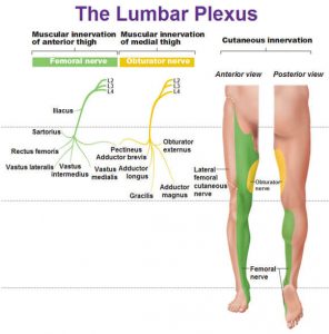
The lumbosacral plexus represents the nerve supply to the lower back, pelvis and legs. Lumbosacral plexopathy is characterized as inflammation and or damage to the nerve bundles in the area of the lumbar and sacral vertebrae regions. Symptoms may include shooting or burning pain, numbness, and decreased movement in the thigh, buttock and/or leg area. It is an uncommon idiopathic disorder(relates to or denotes any disease or condition which arises spontaneously or for which the cause is unknown). It is characterized by the acute onset of severe lower extremity pain followed by wasting and weakness of leg muscles along with variable sensory loss.

To Summarise : Lower Lumbosacral Plexopathy

It predominantly affect the L4–S3 nerve fibers.

There patient complains of a deep boring pain in the pelvis that can radiate posteriorly into the thigh and down into the posterior and lateral calf.

The ankle reflex may be depressed or absent.

Sensory symptoms and signs may be severely deminished over the posterior thigh and posterior-lateral calf and in the foot.

Other Causes of Lumbosacral Plexopathy

Structural
Retroperitoneal hemorrhage (anticoagulation, hemophilia)
Pelvic or abdominal tumor
Aneurysm (common or internal iliac artery)
Endometriosis
Trauma
Nonstructural
Inflammatory (plexitis)
Infarction
Postpartum [during childbirth]
Diabetes (diabetic amyotrophy – most common non-structural cause)
Radiation
Postsurgical (retractor injury)

Please note that the presentation of Lumbosacral Plexopathy has subtle differences to sciatica and discogenic pain, the later conditions being much more common than Lumbosacral Plexopathy.


Physiotherapists in Tralee  Phone 086-7700191 to discuss your condition or to make an appointment.

 Hammer Toes

Hammer toes and mallet toes are foot deformities that occur due to an imbalance in the muscles, tendons or ligaments that normally hold the toe straight. This is a deformity that causes your toe to bend or curl downward instead of pointing forward. It can affect any toe.

Hammer ToesHammer Toes

Causes of Hammer Toes

Hammertoe and mallet toe have been linked to:

  • An imbalance in toe muscles leading to instability, which in turn can cause the toe to contract.
  • Genetics(say one of your parents has it). It may be present at birth.
  • It may develop over time due to arthritis. Diabetes could also increase your risk.
  • An injury in which you stub, jam or break a toe can make it more likely for that digit to develop hammertoe.
  • Poorly fitting and/or high-heeled shoes or footwear that are too tight at the toes crowding your toes into a space in which they can’t lie flat.
  • toe length(if your second toe is too long)
  • Sex (women seem more prone but maybe that is down to fashion ie. high heels etc.?)

Complications of Hammer Toes

At first, a hammertoe or mallet toe might maintain its flexibility. Eventually, the tendons of the toe can contract and tighten, causing your toe to become permanently bent. The toe may become painful and moving it may also be difficult or painful. The raised portion of the toe or toes can rub against your shoes, causing painful corns or calluses.

Prevention of Hammer Toes

Relieving the pain and pressure of hammertoe may involve changing your footwear and/or wearing orthotics. If you have a more severe case of hammertoe or mallet toe, you might need surgery to get relief.

You can avoid many foot, heel and ankle problems with shoes that fit properly. Here’s what to look for when buying shoes:

  • Make sure there is adequate toe room. Avoid shoes with pointed toes.
  • Low heels. Avoiding high heels.
  • Make sure shoes are supportive and comfortable. Lace them up properly

These additional tips can help you buy the right shoes:

  • Buy shoes at the end of the day. Your feet swell slightly throughout the day.
  • Check your size. As you age, your shoe size might change — especially the width. Measure both feet and buy for the larger foot.
  • Buy shoes that fit. Be sure shoes are comfortable before you buy them. If necessary, a shoe repair store might be able to stretch shoes in tight spots, but it’s better to buy them to fit.

For more on hammer toes  see this video

Physiotherapy clinics in Tralee and Dingle phone Eddie on 086-7700191

Iliotibial band syndrome. The I.T. band is not the problem.

iliotibial band syndrome

Regularly I come across  people receiving seemingly endless treatment sessions iliotibial band syndrome, with the main focus of the treatment being to try and loosen out the I.T. band. Below are a few things I believe you should consider when  treating  iliotibial band syndrome.

1. A client is told they have certain weak muscle groups which are causing the problem and are given a load of exercises to correct these weaknesses. Sometimes they are told that they need orthotics. Sometimes these treatment protocols may be necessary, but more often this is just an overuse injury that is being made way too complicated. Say you have been running for a year(or years) with no issues, why are all these ”weaknesses” just now causing you problems? Most likely they are not the cause.  Why do you suddenly need orthotics. Did you change your feet(joke)? It is more likely you are overdoing it a bit in your running.

2. All the emphasis is often placed on the I.T. band and treatment of same. The I.T. band is a long tendon and is a fairly inflexible structure, so concentrating on loosening out that is not going to give much relief in itself. Now if you shift the focus to loosening out the tightened outer quad muscles which attach to the I.T. band by fascia(connective, supportive sheet of tissue, kind of like a very strong clingfilm) and as a result pull on the I.T. band, you will generally start to recover fairly quickly. There is nearly always a section of the outer quad about a quarter of the way up from the knee which will be tight and ropy in this injury. Also don’t forget the Tensor fascia lata muscle. This is up near the hip. It is the muscle that attaches directly to the I.T. band. If this has tightened it also needs to be worked out and loosened also. The I.T band should not be totally ignored of course. Instrument assisted soft tissue release can be excellent in loosening it somewhat, as well as for loosening out tightened quad muscles and fascia.

3. Another problem with this condition is a lack of understanding as to what is causing the pain at the side of the knee. The tension and resulting friction on the I.T. band causes the bursa under it at the side of the knee to become inflamed. This is what causes the pain. Treatment options here include a combination of non steroidal anti-inflammatories, compression with ice(very effective), maybe some electro-acupuncture. You want to compress the icepack into the side of the knee to flatten the inflamed bursa and reduce inflammation. A packet of peas or one of those gel packs will not be cold enough.

4. Rest is needed. This is an overuse injury, remember, but the better and more effective the treatment, the faster the recovery.

Above is not necessarily stuff you will find in a text book but comes from knowledge of anatomy and experience in  successfully treating this condition hundreds of times over the years, often in about two treatment sessions. Injuries are often over-dramatized. Sometimes you just need a little rest, with the right physio treatment protocol to aid recovery.

Muscle twitches

Muscle TwitchesMuscles are made up of fibers that your nerves control. Muscle twitching is caused by minor muscle contraction in the area, or uncontrollable twitching of a muscle group that is served by a single motor nerve fiber. Stimulation or damage to a nerve may cause your muscle fibers to twitch. Most muscle twitches are minor and aren’t usually a cause for concern. Some are common and normal. Others are signs of a nervous system disorder.


Causes

These may include:

  • Autoimmune disorders such as Isaac syndrome.
  • Drug overdose (caffeine, amphetamines, or other stimulants).
  • Lack of sleep.
  • Drug side effect (such as from diuretics, corticosteroids, or estrogens).
  • Exercise (twitching is seen after exercise).
  • Lack of nutrients in the diet (deficiency).
  • Stress.
  • Medical conditions that cause metabolic disorders, including low potassium, and kidney disease, and uremia.
  • Twitches not caused by disease or disorders (benign twitches), often affecting the eyelids, calf, or thumb. These twitches are normal and quite common, and are often triggered by stress or anxiety . These twitches can come and go, and usually do not last for more than a few days.

Nervous system conditions that can cause muscle twitching include:

  • Amyotrophic lateral sclerosis (Lou Gehrig disease)
  • Neuropathy or damage to the nerve that leads to a muscle
  • Spinal muscular atrophy
  • Weak muscles (myopathy)


When to Contact a Medical Professional

Call your health care provider if you have long-term or persistent muscle twitches or if twitching occurs with weakness or loss of muscle.

Physiotherapist in Tralee : Phone 086-7700191

Arterial endofibrosis is an unusual condition, but an important one to keep in mind as a differential diagnosis, especially in the case of cyclists. When an athlete  bends forward and exercises(say like cyclists), it can put pressure on and/or cause a kink in the arteries supplying the working muscles of the leg with oxygen, sugars and so forth.  The artery affected  is usually one of the external iliac arteries which supply the majority of blood to each leg, from the heart.

Arterial Endofibrosis
Fig 1. Arterial Endofibrosis

The theory is that the longer time spent bending forward and exercising, the more the artery can be kinked and irritated. Over time, the body responds to this irritation by making the artery harder, thereby reducing the irritation to the artery (a good outcome), but this hardening also reduces the blood supply to the leg when exercising (a bad outcome for bike racers).

With Arterial endofibrosis the patient typically complains of weakness and cramping, ‘bursting’ or aching pain of a single leg only during heavy efforts on the bike.  They can’t ride through it, and in fact, the the harder they try, the worse it gets.  The onset is usually slow, over months, but extremely consistent. Always with increasing intensity comes decreasing strength.

The patient may get treated by a number of therapists often misdiagnosing the condition, and diagnosing some other exotic sounding condition instead. One thing remains constant though; the symptoms persist. A simple, but not always conclusive test that doctors perform is to exercise the patient, then take their blood pressure at their arm and at their ankle.  In a normal patient these two measurements will be the same or similar.  In a patient with the problem, the blood pressure in the ankle of the effected leg will be lower (because it is getting less blood).

More invasive tests may follow.   Treatment to correct the condition involves surgery. Often it is difficult to confirm the condition with absolute certainty.  This is important because the surgery carries risk, so you really want to be as sure as possible.  Surgery involves cutting out the kinked bit and sewing in another bit of vein from your leg instead, referred to as a ”patch”. Because a vein is a bit ‘flimsier’ than an artery, it bends a bit better. An artery has a muscular wall – which is why it pulses, making a patch using a piece of artery less suitable. Also you can ‘spare’ a bit of vein more than you can spare artery, and as a rule arteries run deep and are harder to harvest.

Return to cycling needs to be managed carefully post surgery, and has to be done under close medical supervision. However, the cyclist can eventually resume normal training/competition etc.

For more information see this video


Physiotherapists in Tralee. Open early until late. Phone 086-7700191.

Bone Spurs – Causes?

Bone spurs(also called osteophytes) are outgrowths of bone occurring along the edge of a bone. Bone spurs are most commonly found in joints, where two bones come together. They also occur close to where muscles, ligaments, or tendons attached to bone. Some common parts of the body affected include the neck (cervical spine), shoulders, hips, hands and heel bone.

Bone spurs typically occur because of continued stress or rubbing on a bone over a prolonged period of time. This can occur due to inflammatory conditions  such as osteoarthritis , tendinitis or tenosynovitis. Normally there is a smooth layer of cartilage on the edges of bones where they come together to form a joint. With osteoarthritis this cartilage layer becomes worn away. This causes the exposed bones to rub against each other. New bone forms in response to this stress and the resulting inflammation. Bone spurs usually develop in areas near tendons and ligaments due to chronic inflammation in these areas. This inflammation can result from friction between these tissues and bone, or from overuse. The bone spur development is the bodies way of trying to protect itself.

Signs and Symptoms

Bone Spurs
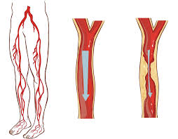
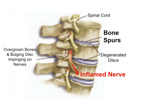
Bone spurs may or may not cause symptoms. Symptoms are location dependent. If bone spurs rub against other bones at joints they can cause pain and/or more limited movement in these joints. They can also be associated with pain, numbness, tenderness, and /or weakness in areas where  they irritate adjacent tissue structures(muscles, ligaments, bones, nerves etc.). If the bone spur rubs against tendons or ligaments, they can sometimes cause a tear in these tissues over time. This is a common cause of tears in certain rotator cuff tendons .

If bone spurs occur in the spine  they can sometimes pinch the nerves (radiculopathy) or spinal cord(myelopathy). A radiculopathy can cause pain, numbness, tingling, or weakness in the arms or legs depending on the area affected. With a myelopathy there can be pain and problems with balance along with weakness.

Diagnosis

Generally X-Rays, MRIs or CT scans are used to confirm suspected cases. Sometimes an experienced practitioner can feel the spur with their finger depending on the area(i.e. heel spur).

When Is Surgery Appropriate ?

In some cases, if symptoms cannot be controlled with more conservative treatment, surgery could be an option. The goal is to remove the bone spur, allowing a return to normal joint motion, or to remove the pressure on muscles, tendons, ligaments, or nerves.


Physiotherapist Tralee  :  Phone 086-7700191