overtraining syndromeOvertraining syndrome, a common cause of persistent tiredness in sportspeople, seen by physio’s, frequently occurs in athletes who are training for competition or a specific event and train beyond their bodies ability to recover. Athletes often exercise longer and harder so that they can improve. However without adequate rest, recovery and nutrition, these training regimens can backfire, actually decreasing performance. Proper training and conditioning requires a balance between overload and recovery. The terms overtraining, overreaching, overtraining syndrome, burnout, and staleness have all been associated with overtraining syndrome and need to be clarified. Overtraining is excessive training with inadequate recovery. Overtraining has also been linked to glycogen depletion(Costill et al. 1988) when inadequate refuelling of muscles glycogen occurs due to low carbohydrate intake. The sooner carbohydrate is consumed following a bout of exercise, the more effective the replenishment of glycogen stores. Adequate quality protein intake would also be important in the case of strength athletes to help protect against overtraining syndrome. Sufficient iron intake is important for endurance athletes especially women.

The term overreaching describes similar symptoms(fatigue, performance decrements, mood state
changes) but is generally of a more transitory nature and is often utilised by sportspeople/coaches
during a typical training cycle to enhance performance. Intense training, in the short term can result
in a decline in performance; however when incorporated with periods of recovery, a
super-compensation effect may occur, with the sportsperson exhibiting enhanced performance
when compared with baseline level (Halson and Jeukendrup, 2004). Overtraining syndrome
develops when there is failed adaptation to overload training due to inadequate regeneration.
Unfortunately , many sportspeople and coaches, especially at amateur level, react to impaired
performance by increasing the intensity of training. This leads to further impairment of
performance, which may , in turn, result in the sportsperson increasing training even further. A
vicious cycle develops which leads overtraining syndrome.

Changes within the central nervous system seem to play an important role in the development of
chronic fatigue and many of the other common signs and symptoms that are frequently seen in
overtraining syndrome, such as disrupted sleep, changes in appetite and weight, irritability, impaired
concentration, decreased motivation, and depressed mood. It has been suggested that alterations in
levels of brain neurotransmitters(e.g. a reduction in serotonin levels) and an increased release of
inflammatory mediators(e.g. cytokines) are important factors in the development of overtraining
syndrome(Anish, 2005). Many of the signs and symptoms that characterize overtraining syndrome are remarkably similar to those of clinical depression. Unfortunately, no single test can detect overtraining in the sportsperson. Probably the simplest and most effective means of monitoring overtraining is self analysis by sportspeople themselves. Daily documentation should include sources and ratings of stress, fatigue, muscle soreness, quality of sleep, irritability, and perceived exertion during training or standardized exercise.

Blood parameters such as red and white blood cell counts, haemoglobin, hematocrit, urea, and
ammonia are usually normal during overtraining. Changes in exercise blood lactate concentration
and blood lactate threshold however have been shown to be good indicators of overtraining but are
influenced by many other factors and are probably only useful if assessed repeatedly.
The initial symptom of the overtraining syndrome is usually fatigue but in time, other symptoms
develop.(Mackinnon and Hooper, 2000) Indicators of overtraining include; decreased performance
despite continued training, persistent fatigue, increased early morning heart rate or resting blood
pressure, frequent illnesses such as upper respiratory tract infections, persistent muscle soreness,
loss of body mass, mood changes, loss of appetite, sleep disturbance, high self-reported stress
levels, irritability, depression , decreased maximal heart rate. Overtraining syndrome can also leave
an athlete more susceptible to injury.

Deep tissue massage and injury prevention advice is where or physiotherapists come in. Feel free to set up an appointment at our Tralee clinic to discuss things. For more details click here.

REFERENCES

Anish, E.J. Exercise and its effects on the central nervous system. Curr. Sports Med. Rep. 2005:4:18-23.

Costill, D.L., Flynn, M.G., Kirwan, J.P. et al. Effects of repeated days of intensified training on muscle glycogen and swimming performance. Med. Sci. Sports Exerc. 1988;20:249-54.

Halson, S.L., Jeukendrup, A.E.https://pubmed.ncbi.nlm.nih.gov/15571428/ Does overtraining exist? An analysis of overreaching and overtraining research. Sports Med. 2004;34(14):967-81.

Mackinnon, L.T., Hooper, S.L. Overtraining and overrreaching: causes, effects, and prevention. In :
Garret, W.E., Kirkendall, D.T., eds. Exercise and Sports Science. Philadelphia : Lippincott, William &
Wilkins, 2000.

Plantar plate injuries are easily missed, probably because a lot of people don’t know what the plantar plate is. Quite they are diagnosed under the general term metatarsalgia. The plantar plate is a deep fibrocartilaginous structure that originates from the metatarsal head and attaches to the proximal phalanx through the joint capsule within the forefoot. Its role is to help stabilize the metatarsophalangeal joints (MTPJ), along with a couple of other structures. The plantar plate also acts as an attachment site for the plantar fascia, so if you load the foot, the medial arch lengthens, the plantar fascia tightens, this engages the plantar plate to plantarflex the proximal phalanx until the toe reaches the ground. This is a simplification of a complex process and is commonly known as the The ‘reversed’ windlass mechanism (with weight-bearing the longitudinal arch flattens, the foot lengthens, the plantar fascia tightens, the proximal phalanx becomes plantarflexed and the mechanism comes to a stop when the proximal phalanx presses against the ground).

plantar plate injury


What causes a plantar plate injury and how common are they?

There are many contributing factors. The first is any activity that exposes the MTPJ to repetitive and excessive dorsiflexion, so think about jumping and running especially in forefoot runners. There are a few biomechanical conditions that increase the load through the plantar plate such as hallux valgus (bunions). As the function through the 1st MTPJ(big toe) is reduced, then we get what is known as low gear propulsion and increased loading through lesser MTPJs, typically the 2nd, 1st, then 3rd and so on. Another condition like having say an irregular metatarsal length, for example, if you have a long 3rd metatarsal, can expose the plantar plate to increased load, as can external factors like high heels. Basically anything that will result in excessive dorsiflexion or ground reaction forces at the MTPJs may increase plantar plate loading.


How does a plantar plate injury present?..

  • The patient will complain of pain on the dorsal and plantar aspects of the MTPJ, usually described as an ache or bruising.
  • Mild oedema may be present along with an episode of trauma, however, trauma is not essential as plantar plate injuries are typically a chronic overuse injury
  • Weight-bearing activities increase pain  – especially dancing, forefoot running, barefoot walking, etc
  • Rest /non-weight bearing reduces pain.
  • High heels or flexible footwear increases pain
  • Reduced plantarflexion strength – The ‘Digital Purchase’ test
    • A quick way to do this, put a piece of paper under the apex of the affected toe and ask the patient to try and stop you pulling the paper away, in a plantar plate injury you will notice the paper is pulled away much more easily.
  • Pain, oedema and positive Digital Lachmans (Anterior Draw) / Vertical Stress.
  • Floating toe, if late-stage hammertoe, or Churchill sign may be present.


Diagnosis of plantar plate injuries


Digital Lachmans / Vertical Stress Test (Fig 1)

Same style of test to assess ACL tears, helps to assess the integrity of the plantar plate, it is quick, easy and a simple test to perform. Stabilise the head of the metatarsal with one hand, using the other hand stabilise the base of the proximal phalanx, apply a vertical force, we are looking for pain and any translocation, it is important to remember this is different from dorsiflexion of the digit.

Fig. 1


There are 2 scoring systems one by Thompson and Hamilton and the other Yu and Judge

Thompson and Hamilton

  • Stage 0, there is no dorsal translocation present of the proximal phalanx.
  • Stage 1 the base of the phalanx, will not dislocate, however, may sublux
  • Stage 2 the base of the phalanx can be dislocated.
  • Stage 3 the phalanx base is in a fixed dislocated position

Yu and Judge

  • Stage 1 mild odema on the plantar MTPJ with dorsal odema often present as well. Tenderness is present on palpation, however no anatomical malalignment.
  • Stage 2 moderate odema is present with a noticeable deviation.
  • Stage 3 odema present around the entire MTPJ with deviation and possible dislocation/subluxation, the odema will reduce however the deformities will remain.

I think the best way to describe the 2 different methods of testing, would be that the Thompson and Hamilton test best describes the integrity of the plantar plate at any given time, whereas the Yu and Judge test describes different stages based on clinical findings on the time of examination.

MRIs, X-rays and Ultrasound

There is still some debate as to whether an MRI scan or ultrasound scan is best for detecting plantar plate injuries. As we know ultrasound is cheaper, however, it is user-dependent, whereas MRI scan is more expensive but we can also get an overall picture of the structures within that area as well. X-ray in weight-bearing (lateral or oblique views) will show subluxation dorsally of the proximal phalanx on the metatarsal head, an anterior-posterior view will show a transverse deformity as well. An x-ray will also rule out other bony pathologies.12


Treatment of plantar plate injuries

The aim of treatments, like most musculoskeletal pathologies, is about managing the load. Essentially we want to try and reduce the ground reaction forces under the affected metatarsal head and reduce the plantarflexion moment of the metatarsal and the dorsiflexion of the phalanx.

Treatment protocols include

  • No barefoot walking/activity modification
  • Footwear advice / Air cast boot – we want to look at using a stiff-soled shoe, or reducing the heel height of a shoe, so footwear like high heels and the flexible minimalist type shoes tend to aggravate a plantar plate injury, the same goes for open-toe shoes and flip-flops, as you must claw your toes to keep these on which again increases the ground reaction force underneath the metatarsal.
  • Stretching / Strengthening – thinking about the mechanics of the foot, if there is tightness within the calf muscles, in turn, could result in early and increased loading through the forefoot, and if you are unable to get adequate dorsiflexion due to calf tightness, then the foot may pronate to compensate for this, which in turn could increase the loading through the lesser MTPJ’s. It is important also to work on strengthening the muscles within the foot.
  • Strapping can be very helpful in reducing pain, using a rigid zinc oxide tape and pulling the toe into a plantarflexed position to help offload a plantar plate (Fig 2).
Fig. 2 Strapping for plantar plate injury


Orthotics

Orthotics can be a useful way to help offload the affected plantar plate. One of the best ways to treat Plantar Plate Injures with or without surgery is using an orthotic device that places the pressure into the archway and off of the ball of the foot. If manufactured and molded correctly, they can keep the tension off the injury and pressure when standing and walking. Combining the orthoses with taping and footwear advice can be quite an effective way of offloading the affected plantar plate, whilst the patient reduces sporting activities.


Steroid injections

Steroid injections can be tried , however repeated intra-articular injections has been shown to result in dislocation of the MTPJ. It has also been suggested that injections into a ligament resulted in destruction of fibrocytes and reduction in tensile strength for up to 1 year which in turn may result in further damage a possible rupture.

A recent case study showing a patient with a plantar plate tear was managed using conservative measures, consisting of taping, activity modification and the use of a Darco boot over a 6 month period, and progressing to stiffed shoe and orthoses and stopped taping. At the 1 year mark, the patient was pain-free with no toe deformity, and on MRI the plantar plate has healed.

So what’s my treatment plan?

  • No barefoot walking for 6 weeks (minimum)
  • To wear stiff-soled shoes
  • Strapping of digit changing every 72 hours
  • Activity modification
  • Orthoses as described as above, plus any other modifications required
  • Stretching and Strength work – Distal and proximal


If conservative measures fail, then it may require referral to a surgeon.


Physiotherapist in Tralee. Ring to discuss your condition, to get a second opinion or to make an appointment. Click here for website.

Herniated Disc vs Bulging Disc

herniated disc
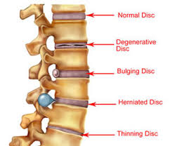
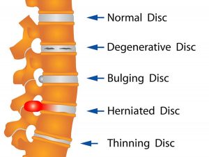
Your spine is made up of lots of bones called vertebrae and discs stacked on top of one another, forming the spinal canal. The nerves of the spinal cord run down the length of the spinal canal. The discs in act like shock absorbers between the vertebrae. These discs are made of two components: a softer center (called nucleus pulposus) surrounded by a tough elastic-like band (called annulus fibrosus). A bulging disc is like a squashed jam doughnut before the jam bursts out. The disc sags and looks like it is bulging outward. With a herniated disc, the outer covering of the disc has a hole or tear. This causes the nucleus pulposus (jelly-like center of the disc) to leak into the spinal canal. It is like the jam leaking out from the inside of a squashed jam doughnut.

Bulging discs are usually caused by age-related degeneration. There is usually a progressive, gradual onset of symptoms. Bulging discs and herniated discs can occur anywhere along the spine. Pain from a herniated disc usually comes on abruptly often affecting one individual nerve root. Herniated discs are often caused by an acute injury. In some cases, you may know the cause of the injury, such as twisting incorrectly.

Herniated and bulging discs are the most common causes of lower back pain, as well as leg pain or ”sciatica.” Between 60% and 80% of people will experience low back pain at some point their lives. Although a herniated disk can be very painful, most people feel much better with just a few weeks or months of nonsurgical treatment, such as using  muscle relaxants and anti-inflammatory medications, along with physiotherapy.  Long term management of these conditions needs to including properly prescribed strengthening and flexibility exercises in order to avoid surgery. Surgery is the final option.

For more on herniated and bulging discs, see video.


Physio in Tralee. Phone 0867700191 to discuss your condition or make an appointment.

Posterior ankle impingement physiotherapyPosterior ankle impingement is a condition characterised by tissue damage at the back of
the ankle joint due to compression of these structures. This occurs when the foot and ankle
are pointed maximally away from the body (plantarflexion – figure 1. ). It may occur when
compressive forces are too repetitive and/or too forceful. This can occurs in the presence of
ankle swelling or bony anomalies, such as additional bone, a condition known as an “os
trigonum”. Posterior ankle impingement is most commonly found in gymnasts, ballet
dancers, and footballers, because they regularly maximally plantarflex their ankles during
their activities. The condition can also occur due to inadequate rehabilitation of an acute
ankle injury (ie. ankle sprain).

Mechanism of Injury

Posterior ankle impingement may develop due to an acute traumatic plantar hyperflexion
event, such as an ankle sprain. It may also occur as a result of repetitive low-grade trauma
associated with plantar hyperflexion, say like in case of a female ballet dancer. It is
important to differentiate between these two, because the latter, that is posterior
impingement from overuse, has a better prognosis.
The anatomy of the posterior ankle is a key factor in the occurrence of posterior
impingement syndrome . The more common causes of the condition are osseous in nature,
such as the os trigonum, an elongated posterolateral tubercle of the talus (known as
Stieda’s process), a downward sloping posterior lip of the tibia, an osteophyte from the
posterior distal tibia , or a prominent posterior process of the calcaneus. However, posterior
impingement can also be soft tissue related, as with a thickened posterior joint capsule ,
post-traumatic scar tissue, post-traumatic calcifications of the posterior joint capsule, or
loose bodies in the posterior part of the ankle joint. Symptoms for all of these conditions
relate to physical impingement of osseous or soft tissue structures, resulting in painful
limitation of the full range of ankle movement.
The most common cause ''os trigonum'' is an extra (accessory) bone that sometimes
develops behind the ankle bone (talus). The mineralized os trigonum appears between the
ages of 7 and 13 years and usually fuses with the talus within 1 year, forming the trigonal
(Stieda) process. It may remain as a separate ossicle in 7-14% of patients, and is often
bilateral(in both ankles). An os trigonum can be a focus of osseous abutment against other
structures. Pain can also be caused by disruption of the cartilaginous synchondrosis
between the os trigonum and the lateral talar tubercle as a result of repetitive microtrauma
and chronic inflammation.
In the case of soft tissue impingement it usually results from scarring and fibrosis associated
with synovial, capsular, or ligamentous injury ie. bad ankle sprain. It is thought that this
type of manifestation usually usually occurs when a significant soft-tissue component
forms. The soft-tissue component can consist of synovial thickening throughout the
posterior capsule or be more focal, involving the posterior intermalleolar or talofibular ligament. The flexor hallucis longus tendon runs in the groove between the lateral and
medial processes of the talus and can also be injured in posterior impingement, resulting in
tenosynovitis.


Signs and symptoms

Patients who have posterior impingement complain of chronic deep posterior ankle pain
worsened by forced plantar flexion or push-off forces as occur during activities such as
ballet dancing, jumping, or running downhill. In some patients, forced dorsiflexion(opposite
to plantarflexion) is also painful. Physical examination reveals pain on palpation over the posterolateral talar process, which is located along the posterolateral aspect of the ankle between the Achilles and peroneal
tendons . Passive forced plantar flexion results in pain and often a grinding
sensation as the posterolateral talar process is entrapped between the posterior tibia and
calcaneus.


Diagnosis of posterior ankle impingement

A thorough examination by an experienced practitioner may be all that is necessary to
diagnose posterior ankle impingement. Further investigations such as an X-ray, MRI, CT scan
or Ultrasound may help confirm diagnosis.


Physiotherapist in Tralee, Co. Kerry………..Phone 0867700191 to make an appointment or discuss your condition.

Everyone’s spine has curves, from your neck down to your lower back. These curves, which create the spine’s ”S” shape, are  lordotic in shape in the neck and lower back, and  kyphotic in shape for the upper back. They help your body 1. absorb shock,  2. support the weight of the head, 3. align your head over your pelvis, 4. stabilize and maintain its structure, 5. move and bend flexibly. When these curves are exaggerated through poor posture or hereditary conditions like scoliosis, spinal pain and problems can develop over time.


Kyphosis

Kyphosis
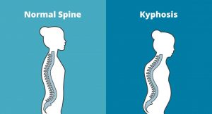
Kyphosis is an exaggerated, forward rounding of the back. It can occur at any age but is more common in older people. Age-related kyphosis is often due to weakness in the spinal bones that causes them to compress or crack. Other types of kyphosis can appear in infants or teens due to malformation of the spine or wedging of the spinal bones over time. Mild kyphosis causes few problems. Severe kyphosis can cause pain and be disfigurment. Treatment for kyphosis depends on your age, and both the cause and the effects of the curvature.


Lordosis

Lordosis
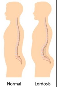
Some curvature in the lower back is normal. However, if your curve arches too far inward, it’s called lordosis, or swayback. Lordosis can affect your lower back and/or neck. The condition can lead to excess pressure on the spine, causing pain and discomfort. It can affect your ability to move if it’s severe and left untreated. Treatment of lordosis depends on how serious the curve is and how you got lordosis. There’s little medical concern if your lower back curve reverses itself when you bend forward. You can probably manage your condition with physical therapy and daily exercises.


Scoliosis

scoliosis
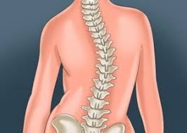
Scoliosis is a medical condition in which a person’s spine curves sideways, usually in an  “S” or “C” shape. In some, the degree of curvature  remains stable, while in others, it increases over time. Mild scoliosis does not typically cause problems, however severe cases can interfere with breathing. The cause of most cases is unknown, but is believed to involve a combination of genetic and environmental factors. Diagnosis is confirmed by Xray. Treatment depends on the degree of curve, location, and cause.

Minor curves may simply be watched periodically. Treatments may include bracing or surgery in more severe cases. The brace must be fitted to the person and used daily until growing stops. Evidence that chiropractic manipulation dietary supplements, or exercises can prevent the condition from worsening is non existent. However, exercise is still recommended due to its other health benefits. Scoliosis occurs in about 3% of people. It most commonly occurs between the ages of 10 and 20. Girls typically are more severely affected than boys.


Physiotherapists in Tralee Phone 086-7700191

Arthritis – Rheumatoid vs Osteoarthritis


Rheumatoid arthritis

rheumatoid arthritis
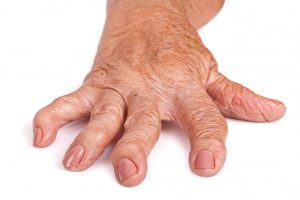
Rheumatoid arthritis is a chronic inflammatory disorder that can affect more than just your joints. It is an autoimmune disease occurring when your immune system mistakenly attacks the  synovium , the lining of the membranes that surround your joints. The resulting inflammation causes the synovium to thicken, eventually destroying the cartilage and bone within the joint. The tendons and ligaments holding the joint together also weaken and stretch. Gradually, the joint loses its shape and alignment.

It is unknown exactly  what starts the process, although a genetic component appears likely.  Newly developed  medications have greatly improved treatment options.  However, severe rheumatoid arthritis can still cause physical disabilities.

 Symptoms

Signs and symptoms of rheumatoid arthritis may include:

  • Tender, warm, swollen joints
  • Joint stiffness that is usually worse in the mornings and after inactivity
  • Fatigue, fever and weight loss

Early rheumatoid arthritis tends to affect your smaller joints first — particularly the joints of the fingers and toes. As the disease progresses, symptoms often spread to the wrists, knees, ankles, elbows, hips and shoulders. In most cases, symptoms occur in the same joints on both sides of your body.

Rheumatoid arthritis can also affect many non joint structures. This is the case in about 40 per cent of sufferers. These include:

  • Skin
  • Eyes
  • Lungs
  • Heart
  • Kidneys
  • Salivary glands
  • Nerve tissue
  • Bone marrow
  • Blood vessels

Rheumatoid arthritis signs and symptoms can vary in severity and may come and go. Periods of increased disease activity, called flare ups, alternate with periods of relative remission — when the swelling and pain fade or disappear.

Risk factors

  • Your sex : Women are more prone than men .
  • Age : Can occur at any age, but most common between the ages of 40 and 60.
  • Family history : Increased risk if there is a family history of the disease.
  • Smoking : Cigarette smoking increases your risk of developing rheumatoid arthritis.
  • Obesity: People who are overweight or obese appear to be at somewhat higher risk .

Diagnosis and Blood tests

People with rheumatoid arthritis often have an elevated erythrocyte sedimentation rate (ESR, or sed rate) or C-reactive protein (CRP), which may indicate the presence of an inflammatory process in the body. Other common blood tests look for rheumatoid factor and anti-cyclic citrullinated peptide (anti-CCP) antibodies. Rheumatoid arthritis can be difficult to diagnose in its early stages because the early signs and symptoms mimic those of many other diseases. There is no one blood test or physical finding alone that confirms diagnosis.

Imaging tests

Your doctor may recommend X-rays to help track the progression of rheumatoid arthritis in your joints over time. MRI and ultrasound tests can help your doctor judge the severity of the disease in your body.

Treatment

There is no cure for rheumatoid arthritis. Recent studies indicate that remission of symptoms is more likely when treatment begins early using medications known as disease-modifying antirheumatic drugs (DMARDs).

The types of medications recommended by your doctor will depend on the severity of your symptoms and how long you’ve had the rheumatoid arthritis. Medications used include :

  • NSAIDs : Nonsteroidal anti-inflammatory drugs (NSAIDs) can relieve pain and reduce inflammation.
  • Steroids : Corticosteroid medications, such as prednisone, reduce inflammation and pain and slow joint damage.
  • Disease-modifying antirheumatic drugs (DMARDs) : These drugs can slow the progression of rheumatoid arthritis and save the joints and other tissues from permanent damage. Common DMARDs include methotrexate (Trexall, Otrexup, Rasuvo), leflunomide (Arava), hydroxychloroquine (Plaquenil) and sulfasalazine (Azulfidine).
  • Biologic agents : Also known as biologic response modifiers. These are a newer class of DMARDs(Disease-modifying antirheumatic drugs) .These drugs can target parts of the immune system that trigger inflammation that causes joint and tissue damage. Biologic DMARDs are usually most effective when paired with a nonbiologic DMARD, such as methotrexate.

All the above drugs have side effects which need to be monitored.

Surgery

Medications can slow joint damage due to rheumatoid arthritis. When the damage becomes excessive surgery may need to be considered to repair the damaged joints.

Osteoarthritis

Osteoarthritis is the most common form of arthritis. It occurs when the cartilage that cushions the ends of bones in your joints gradually deteriorates. Cartilage is a firm, slippery tissue that permits nearly frictionless joint motion. In osteoarthritis, the smooth surface of the cartilage becomes roughed and worn. Eventually, if the cartilage wears down completely, you may be left with bone rubbing on bone. The disorder most commonly affects joints of the hands, knees, hips and spine.

Osteoarthritis symptoms can usually be effectively managed, although the underlying process cannot be reversed. Staying active, maintaining a healthy weight and other treatments may slow progression of the disease and help improve pain and joint function. When joint pain and damage is severe, doctors may suggest joint replacement surgery.

 Symptoms

Osteoarthritis symptoms often develop slowly  over time. Signs and symptoms may include:

  • Pain. Your joint may hurt during or after movement.
  • Tenderness. Your joint may feel tender when you apply light pressure to it.
  • Stiffness. Joint stiffness may be most noticeable when you wake up in the morning or after a period of inactivity.
  • Loss of flexibility. You may not be able to move your joint through its full range of motion.
  • Grating sensation. You may hear or feel a grating sensation when you use the joint.


Risk factors

Factors that may increase your risk of osteoarthritis include:

  • Older age. The risk of osteoarthritis increases with age as joints wear over time.
  • Sex. Women are more likely to develop osteoarthritis, though it isn’t clear why.
  • Obesity. Carrying extra body weight contributes to osteoarthritis in several ways, and the more you weigh, the greater your risk. Increased weight puts added stress on weight-bearing joints, such as your hips and knees. In addition, fat tissue produces proteins that may cause harmful inflammation in and around your joints.
  • Joint injuries. Injuries, such as those that occur when playing sports or from an accident, may increase the risk of osteoarthritis.
  • Certain occupations. If your job includes tasks that place repetitive stress on a particular joint, that joint may eventually develop osteoarthritis.
  • Genetics. Some people inherit a tendency to develop osteoarthritis.
  • Bone deformities. Some people are born with malformed joints or defective cartilage, which can increase the risk of osteoarthritis.


Summary

Rheumatoid arthritis is an auto-immune response where your body mistakenly attacks its own joints. Osteoarthritis is more or less due solely to wear on a particular joint over time.


Aside

Sometimes MRI results or Xray results  state there is osteoarthritis present. With age most of us have some level of osteoarthritis due to wear on our joints. The important thing which your doctor will discuss with you is whether this level is normal for your age. A person can have mild osteoarthritis with no pain or symptoms and may have nothing to worry about.


Physiotherapist Tralee phone 086-7700191

Natural Anti-inflammatoriesNatural Anti-inflammatories

There are plenty advertisements for supplements like glucosamine and chondroitin claiming they will reduce inflammation and even rebuild cartilage etc. Most of these supplements have absolutely no evidence to back their claims, or they promote a few poorly carried out studies sponsored by the manufacturers. Three foods with quiet an amount of published scientific studies substantiating their usefulness as natural anti-inflammatories are turmeric, ginger and cinnamon.

Turmeric is a brilliant yellow spice commonly used in Indian cuisine and found in any grocery store. It is the curcumin in turmeric that has the anti-inflammatory properties.  You need a small amount of black pepper and fat in your food also to help with its absorption. Ginger is a zesty spice used in many cuisines. You can buy it powdered or as a fresh root in most supermarkets. Cinnamon is a popular spice often used to flavor baked treats. It is  better to use ceylon cinnamon rather than cassia cinnamon as the casia version is higher in toxins. These three spices have been used as medicines for centuries .  You can use all three(ginger, turmeric, cinnamon) in powder form, preferably organic.

Here is a little recipe that I got from the internet  and it actually  tastes quiet pleasant. Turmeric can be hard to eat unless it is hidden in a curry or a soup or something like that to mask the earthy flavor, but this recipe softens its taste.

1 cup of warm whole fat milk

1 teaspoon of turmeric powder

half teaspoon of ginger powder

half teaspoon of cinnamon powder

1 to 2 dessertspoons of honey

good pinch of black pepper

Blend it all up and drink. You would need to drink this daily to reduce inflammation.

Do the research online yourself and make up your own mind. Don’t use ”Google” as this fires everything at you both true and untrue. Key in ”Google Scholar”. This brings up a sub-site of Google which is more evidence based and shows all the scientific publications on your search.


Physiotherapy in Tralee – Phone 086-7700191

Arterial endofibrosis is an unusual condition, but an important one to keep in mind as a differential diagnosis, especially in the case of cyclists. When an athlete  bends forward and exercises(say like cyclists), it can put pressure on and/or cause a kink in the arteries supplying the working muscles of the leg with oxygen, sugars and so forth.  The artery affected  is usually one of the external iliac arteries which supply the majority of blood to each leg, from the heart.

Arterial Endofibrosis
Fig 1. Arterial Endofibrosis

The theory is that the longer time spent bending forward and exercising, the more the artery can be kinked and irritated. Over time, the body responds to this irritation by making the artery harder, thereby reducing the irritation to the artery (a good outcome), but this hardening also reduces the blood supply to the leg when exercising (a bad outcome for bike racers).

With Arterial endofibrosis the patient typically complains of weakness and cramping, ‘bursting’ or aching pain of a single leg only during heavy efforts on the bike.  They can’t ride through it, and in fact, the the harder they try, the worse it gets.  The onset is usually slow, over months, but extremely consistent. Always with increasing intensity comes decreasing strength.

The patient may get treated by a number of therapists often misdiagnosing the condition, and diagnosing some other exotic sounding condition instead. One thing remains constant though; the symptoms persist. A simple, but not always conclusive test that doctors perform is to exercise the patient, then take their blood pressure at their arm and at their ankle.  In a normal patient these two measurements will be the same or similar.  In a patient with the problem, the blood pressure in the ankle of the effected leg will be lower (because it is getting less blood).

More invasive tests may follow.   Treatment to correct the condition involves surgery. Often it is difficult to confirm the condition with absolute certainty.  This is important because the surgery carries risk, so you really want to be as sure as possible.  Surgery involves cutting out the kinked bit and sewing in another bit of vein from your leg instead, referred to as a ”patch”. Because a vein is a bit ‘flimsier’ than an artery, it bends a bit better. An artery has a muscular wall – which is why it pulses, making a patch using a piece of artery less suitable. Also you can ‘spare’ a bit of vein more than you can spare artery, and as a rule arteries run deep and are harder to harvest.

Return to cycling needs to be managed carefully post surgery, and has to be done under close medical supervision. However, the cyclist can eventually resume normal training/competition etc.

For more information see this video


Physiotherapists in Tralee. Open early until late. Phone 086-7700191.

Muscle Tear or Spasm ? Which is it ?

muscle tear
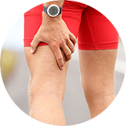
When somebody first presents in clinic with a muscle injury, it is important to first determine whether it is a muscle spasm or a tear. A spasm/cramp can often be worked out in a session with deep tissue massage and maybe some dry needling and stretching, allowing almost immediate turn to training. A muscle tear takes much longer to recover from, the length of time greatly depending on the degree of tearing. Treatment takes longer and a rehabilitation program is also essential.  A muscle spasm is caused by an involuntary contraction of that muscle. It is usually sudden, can cause significant pain and can limit the use of the muscle for a short period of time.

What is a Spasm?

 A muscle spasm is an involuntary contraction of that muscle. It is usually sudden, can cause significant pain and can limit the use of the muscle for a short period of time.

Spasm Causes

Spasms are usually caused by problems such as overworking a muscle, poor hydration, electrolyte imbalances, insufficient blood flow to the muscle or nerve compression. A pulled muscle is actually a tear in the muscle tissue. This occurs when the muscle is strained to the point where the muscle is damaged. The more significant the strain, the more the muscle can be torn.

What is a Muscle pull/Tear?

A pulled muscle is actually a tear in the muscle tissue. This occurs when the muscle is strained to the point where the muscle is damaged. The more significant the strain, the more the muscle can be torn.Pulled muscles are usually the result of not preparing the muscle for work through proper stretching, placing too much tension on the muscle or over-using the muscle. Soreness in the affected muscle is usually the first symptom. Using the muscle will be painful and difficult. In severe pulls, bruising may be present. The affected muscle will become very tender to the touch and some swelling may occur.

Pulled Muscle Causes

Pulled muscles are usually the result of one or a combination of (1) not preparing the muscle for work because of insufficient warmup,(2) lack of a proper stretching program after training to help the muscle recover,(3) placing too much tension on the muscle all of a sudden, (4) or over-using the muscle. Sharp pain in the affected muscle is usually the first symptom. Using the muscle will be painful and difficult. In severe pulls, bruising may be present. The affected muscle will become very tender to the touch and some swelling may occur.

The use of anabolic steroids is frequently linked to severe muscle tears as the body hasn’t had time to adapt to the increased workload . All training needs gradual progression and enough recovery time after training. The higher the intensity in training , the more time needed to recover. So for an athlete, periods of maximal performance require more recovery time.

For treatment of muscle tears or spasms phone 086-7700191. We are physiotherapists in Tralee.


Low Back Pain – Facet vs Disc

Two of the most  common causes of low back pain presenting in clinic are discogenic (disc) and facet joint related pain. Injury to either can cause severe discomfort, limiting a person’s ability to carry out normal daily activities. Facet joints are small articulations along each segment at the back of the spine, and help control the movement of the spine.  There are two of these joints at each vertebral level. There are intervertebral discs between each level  and these act as primary shock absorbers. They are  generally between a quarter and a half inch in height, interconnecting the bodies of the vertebral segments.  It can be considered that the discs and the facets create joint complexes that allow for both shock absorption and movement at each segment of the spine.

Low back pain
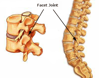
One of the primary symptoms when a  lumbar facet joint locks is an increase in pain on extension of the lower back.  Basically there is more pain when you try and straighten up your lower back or lean back on it. This position loads pressure on the locked lumbar facet joints.   Usually with this type of injury there is pain relief to a certain extent when a person bends forward.  The reason for this is that forward flexion decompresses the facet joint articulations, releasing some of the pressure on the joints and hence the associated pain. There can be nerve irritation with this injury but it is usually more general and not as severe as with discogenic pain.

Low back pain
Disc disorders

When we start talking about disc injury, we are usually referring to a small tear in the outer annular fibers of a disc that has either resulted in a bulge, a protrusion, or an extrusion of the disc particle(see diagram).  Pain occurs when a disc touches off a nerve due to one of these injuries, triggering a series of reactions, some of which include muscle spasm, inflammation and pain.   Often the nerve root irritation sends pain signals down either the front or back of the leg depending on which disc level is affected. In contrast to facet joint injuries, with disc injuries, bending forward causes a significant increase in pain, as it usually increases compression of the disc against the nerve.  So again to generalize, disc injuries are usually more sensitive to forward flexion,  whereby facet injuries are very sensitive to backward extension. A classic and very telling sign of a disc injury is where the spine becomes visibly curved off to one side as the disc tries to get away from the nerve it is touching. You will visibly see the person’s spine twisted off to one side.

With a locked facet joint, a simple osteopathic manipulation can give a person an immediate 60 – 70 % relief from pain symptoms.  The person is often almost back to normal the next day. With a facet joint injury, you are simply unlocking the locked facet joint in which the limited movement and was causing the problem. With a disc injury, you are trying to take pressure off the disc in the hope that it stops pressing against the nerve. Once off lumbar facet joint manipulation can play it’s part in recovery from disc disorders, if used at the right time by an experienced practitioner. Again this is used to mobilize the area and try and take pressure off the disc.

Often with a suspected disc injury a physio etc. may need to refer a patient to a doctor for a second opinion and most possibly medication( NSAIDs, muscle relaxant, nerve blocker), depending on the level of pain and discomfort. The doctor may also refer the patient for an MRI if they need more information on the level of damage. Physio generally involves deep tissue massage, dry needling, spinal manipulation at the right time. A rehab program involving pilates type exercises and some gentle stretching exercises etc. is paramount once a patient starts to improve. Continuous spinal manipulations several times a week for extended periods pushed by certain professions could do more harm than good, causing sheering of the disc and slowing healing.

”Putting back in a slipped disc” by manipulation is a myth. When somebody gets a manipulation done, the little crack you hear is when the facet joint gaps or unlocks. It is not the disc going back into place. Sometimes unlocking a facet joint in the area, if it has locked up say due to muscle spasm etc. can help take pressure of the disc in the area.

Eddie O Grady Physiotherapy, Tralee, Co. Kerry