Lumbosacral Plexopathy

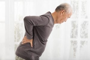
Lumbosacral Plexopathy : Recently I had a patient present with footdrop (foot drops and drags as you walk due to weakness in dorsiflexor muscles), also severe pain down the leg, along with lumbar-sacral pain(lower back area). Initially this might appear to be signs of disc herniation with nerve root compression, even though the footdrop would be fairly uncommon with that condition. An MRI showed the spine to be normal. We also looked at anything that might have injured the peroneal nerve which is quiet close to the skin on the lower leg, which in itself could cause the footdrop, but there was no sign of injury. We were also able to rule out diabetes which can lead to nerve damage.

So where did we go next? Answer : An electromyography (EMG)(nerve conduction study). Electromyography is a diagnostic procedure used to assess the health of muscles and the nerve cells that control them (motor neurons). EMG results can reveal nerve dysfunction, muscle dysfunction or problems with nerve-to-muscle signal transmission. As a result of this testing by a specialist, the client was diagnosed as having a Lumbosacral plexopathy. In this case it seemed that the nerve had been damaged by a viral or bacterial infection. More tests need to be done to confirm and treat this infection.

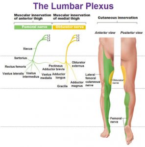
The lumbosacral plexus represents the nerve supply to the lower back, pelvis and legs. Lumbosacral plexopathy is characterized as inflammation and or damage to the nerve bundles in the area of the lumbar and sacral vertebrae regions. Symptoms may include shooting or burning pain, numbness, and decreased movement in the thigh, buttock and/or leg area. It is an uncommon idiopathic disorder(relates to or denotes any disease or condition which arises spontaneously or for which the cause is unknown). It is characterized by the acute onset of severe lower extremity pain followed by wasting and weakness of leg muscles along with variable sensory loss.

To Summarise : Lower Lumbosacral Plexopathy

It predominantly affect the L4–S3 nerve fibers.

There patient complains of a deep boring pain in the pelvis that can radiate posteriorly into the thigh and down into the posterior and lateral calf.

The ankle reflex may be depressed or absent.

Sensory symptoms and signs may be severely deminished over the posterior thigh and posterior-lateral calf and in the foot.

Other Causes of Lumbosacral Plexopathy

Structural
Retroperitoneal hemorrhage (anticoagulation, hemophilia)
Pelvic or abdominal tumor
Aneurysm (common or internal iliac artery)
Endometriosis
Trauma
Nonstructural
Inflammatory (plexitis)
Infarction
Postpartum [during childbirth]
Diabetes (diabetic amyotrophy – most common non-structural cause)
Radiation
Postsurgical (retractor injury)

Please note that the presentation of Lumbosacral Plexopathy has subtle differences to sciatica and discogenic pain, the later conditions being much more common than Lumbosacral Plexopathy.


Physiotherapists in Tralee  Phone 086-7700191 to discuss your condition or to make an appointment.

ROTATOR CUFFRotator Cuff Tears

Your arm is kept in your shoulder socket by your rotator cuff. The rotator cuff consists of four muscles, the supraspinatus muscle, the infraspinatus muscle, the terres minor muscle, and the subscapularis muscle. These muscles attach the humerus to the shoulder blade and help lift and rotate your arm.

Your shoulder is made up of three bones: your upper arm bone (humerus), your shoulder blade (scapula), and your collarbone (clavicle). The shoulder is a ball-and-socket joint: the ball, or head, of your upper arm bone fits into a shallow socket in your shoulder blade.

There is also a lubricating sac called a bursa between the rotator cuff muscles and the bone on top of your shoulder (acromion). The bursa allows the rotator cuff tendons to glide freely when you move your arm. Sometimes it may become inflamed and painful due to overuse.


Rotator Cuff Tears – Causes

There are two main causes of rotator cuff  injury and degeneration.


1. Acute Tears

If you fall down on your outstretched arm or lift something too heavy with a jerking motion, you can tear your rotator cuff at that moment.

2. Degenerative Tears

Many rotator cuff tears are the result of a wearing down of the tendon occurring slowly over time. They are more common in the dominant arm.

Several factors contribute to degenerative, or chronic, rotator cuff tears.

  • Repetitive stress : Repeating the same shoulder motions again and again can stress your rotator cuff muscles and tendons. Baseball, tennis, rowing, and weightlifting are examples of sports activities that can put you at risk for overuse tears. Many jobs and routine chores can cause overuse tears, as well.
  • Lack of blood supply : As we get older, the blood supply to our rotator cuff tendons reduces. Without a good blood supply, the body’s natural ability to repair tendon damage is impaired. This can ultimately lead to a tendon tear.
  • Bone spurs : As we age, bone spurs (bone overgrowths) often develop on the underside of the acromion bone. When we lift our arms, the spurs rub on the rotator cuff tendon. This is part of a condition which comes under the general term of ”shoulder impingement”.  Over time this can damage the tendon, making a tear more likely. The supraspinatus muscle is probably the more common of the rotator cuff muscles to get injured as we age. This is because the supraspinatus passes under the acromion process before attaching to the greater tubercle of the humerus.
  • Poor posture : With poor posture over time the shoulders start to protrude forward making the subacromion space smaller and causing shoulder impingement, sometimes leading to rotator cuff muscle/tendon damage, especially the supraspinatus muscle/tendon.

 Symptoms

The most common symptoms include:

  • Pain at rest and at night, particularly if lying on the affected shoulder
  • Pain when lifting and lowering your arm or with specific movements
  • Weakness when lifting or rotating your arm

Tears that happen suddenly, such as from a fall, usually cause intense pain immediately. There may be a snapping sensation or feeling of something giving way, followed by immediate weakness/severe pain in moving your upper arm.Tears that develop slowly due to overuse also cause pain and arm weakness but the increase in pain intensity occurs over time. You may have pain in the shoulder when you lift your arm, or pain that moves down your arm. At first, the pain may be mild and only present when lifting your arm over your head, such as reaching into a cupboard. Over time, the pain may become more noticeable at rest, and no longer goes away with medications. You may have pain when you lie on the injured side at night. The pain and weakness in the shoulder may make routine activities such as combing your hair or reaching behind your back more difficult.

There are different types of tears.

  • Partial tear : This type of tear is also called an incomplete tear. It damages the tendon, but does not completely sever it.
  • Full-thickness tear : This type of tear is also called a complete tear. It separates all of the tendon from the bone.


Rotator Cuff Tears – Imaging Tests

Magnetic resonance imaging (MRI) or ultrasound are probably the best way to confirm tears, bursitis, tendinopathies or bone spurs in the rotator cuff area.

 Rotator Cuff Tears -Treatment

If you have a partial rotator cuff tear and you keep using it despite increasing pain, you may cause further damage.  A rotator cuff tear can get larger over time.

Chronic shoulder and arm pain are good reasons to see your doctor. Early treatment can prevent your symptoms from getting worse. It will also get you back to your normal routine that much quicker.

The goal of any treatment is to reduce pain and restore function. There are several treatment options, and the best option is different for every person. In planning your treatment, your doctor will consider your age, activity level, general health, and the type of tear you have.

Nonsurgical Treatment

Nonsurgical treatment options may include:

  • Rest. Your doctor may suggest rest and limiting overhead activities. He or she may also prescribe a sling to help protect your shoulder and keep it still.
  • Activity modification. Avoid activities that cause shoulder pain.
  • Nonsteroidal anti-inflammatory medication. Drugs like ibuprofen and naproxen reduce pain and swelling.
  • Strengthening exercises and physical therapy. Specific exercises will restore movement and strengthen your shoulder. Your exercise program will include stretches to improve flexibility and range of motion. Strengthening the muscles that support your shoulder can relieve pain and prevent further injury.
  • Cortisone steroid injection. This is not recommended with a muscle or tendon tear and can actually lead to more damage in the long-term. One exception would be in the case of a subacromial bursitis where it can be very effective in shrinking the bursa back to normal size and reducing the inflammation.

Surgical Treatment

Your doctor may recommend surgery if your pain does not improve with nonsurgical methods. Continued pain is the main indication for surgery. If you are very active and use your arms for overhead work or sports, your doctor may also suggest surgery.

Other signs that surgery may be a good option for you include:

  • Your symptoms have lasted 6 to 12 months
  • You have a large tear
  • You have significant weakness and loss of function in your shoulder
  • Your tear was caused by a recent, acute injury

Surgery to repair a torn rotator cuff most often involves re-attaching the tendon to the head of humerus (upper arm bone).

Home rehab exercises for the rotator cuff muscles click here


Physiotherapist in Tralee : Phone 086-7700191

Common Knee Injuries

1. Chondromalacia Patella

knee injuries
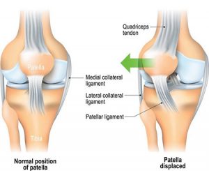
Knee injuries like Chondromalacia patella result from degeneration of cartilage on the underside the kneecap (patella)due to poor alignment of the kneecap (patella) as it slides over the lower end of the thighbone (femur). This condition can also be referred to as patellofemoral syndrome.  It usually presents as pain in the front of the knee aggravated by activities such as running, jumping, climbing or descending stairs etc., or by prolonged sitting with knees in a moderately bent position. Patients with chondromalacia patella frequently have abnormal patellar “tracking” toward the lateral (outer) side of the femur. Some patients may also have a vague sense of “tightness” or “fullness” in the knee area with the condition. Mild swelling of the knee area may also occur.

2. Meniscus TearKnee injuries

knee injuries
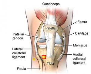
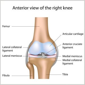
Any activity that causes you to forcefully twist or rotate your knee, especially when putting your full weight on it, can lead to a torn meniscus. Each of your knees has two menisci — C-shaped pieces of cartilage that act like a cushion between your shinbone and your thighbone. A torn meniscus causes pain, swelling and stiffness. You might also feel your knee lock and have trouble extending it fully. Conservative treatment — such as rest, ice and medication — is sometimes enough to relieve the pain of a torn meniscus and allow the injury time to settle down on its own. In other cases, however, a torn meniscus requires surgical repair.

The meniscus weakens with age. Tears are more common in people over the age of 30. If you have osteoarthritis, you’re at higher risk of injuring your knee or tearing your meniscus. When an older person experiences a meniscus tear, it’s more likely to be related to degeneration. This is when the cartilage in the knee becomes weaker and thinner.


3. Cruciate ligament rupture

knee injuries
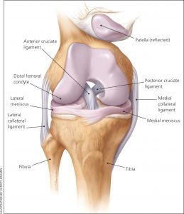
These ligaments are found inside your knee joint. They cross each other to form an “X” with the anterior cruciate ligament in front and the posterior cruciate ligament in back. The cruciate ligaments stabilize the knee. About half of all injuries to the anterior cruciate ligament occur along with damage to other structures of the knee, such as articular cartilage, meniscus, or other ligaments. Partial tears of the anterior cruciate ligament are rare; most ACL injuries are complete or near complete tears.

The  cruciate ligaments can be injured in several ways such as : changing direction rapidly ; Stopping suddenly ; Slowing down while running ; Landing from a jump incorrectly ; Direct contact or collision to the knee.

When you tear a  cruciate ligament, you might hear a “popping” noise and you may feel your knee give way under you. Other typical symptoms include:

  • Pain with swelling. Within 24 hours, your knee will swell. If ignored, the swelling and pain may resolve on its own. However, if you attempt to return to sports, your knee will probably be unstable and you risk causing further damage to the cushioning cartilage (meniscus) of your knee.
  • Loss of full range of motion
  • Tenderness along the joint line
  • Discomfort while walking

Treatment for a cruciate ligament tear varies depending on the patient’s individual needs. For example, the young athlete involved in agility sports will need surgery to return safely to sports. Less active, older individuals may be able to return to a quieter lifestyle without surgery.


4. Collateral ligament strain/ruptureKnee injuries

physiotherapists in DingleThe collateral ligaments of the knee are located one on either side of your knee joint. They help connect the bones of your upper and lower leg, inside your knee joint. The collateral ligaments help keep your knee stable. They keep your leg bones in place and keep your knee from moving too far sideways. A collateral ligament injury occurs when the ligaments are stretched or torn. A partial tear occurs when only part of the ligament is torn. A complete tear occurs when the entire ligament is torn into two pieces. A collateral ligament injury can occur if you get hit very hard on the inside or outside of your knee, or when you have a twisting injury. Sometimes the ligaments just sprain and this is easier to recover from and requires no surgery.

With a collateral ligament injury, you may notice:

  • Your knee is unstable and can shift side to side as if it “gives way”
  • Locking or catching of the knee with movement
  • Knee swelling
  • Knee pain along the inside or outside of your knee

Various orthopedic tests can help diagnose  collateral ligament damage. More detailed knowledge of the injury can be obtained by MRI. Injured ligaments are considered “sprains” and are graded on a severity scale.


If you have a collateral ligament injury, you may need:

  • Crutches to walk until the swelling and pain get better
  • A brace to support and stabilize your knee
  • Physical therapy to help improve joint motion and leg strength

A lot of the time people don’t need surgery for collateral ligament damage  but more serious cases may.


5. Baker’s Cyst – Knee injuries

 knee injuries

Baker’s cyst is a fluid-filled swelling that causes a lump at the back of the knee, leading to tightness and restricted movement. Usually, this condition is due to an underlying problem affecting the knee joint, such as arthritis or a cartilage injury.


6. Patella Tendinopathy

knee injuriesPatellar tendinopathy(also known as Jumper’s knee) is an overuse injury affecting this knee tendon. There are a number of factors which can contribute to the development of patellar tendinopathies. These include:

  • Rapid increase in amount of training
  • Sudden increase in training intensity
  • Playing/training on rigid surfaces
  • Tight quadriceps and hamstring muscles
  • Poor foot posture, knee or hip control.

Tendinopathies usually causes pain, stiffness, and loss of strength in the affected area. The pain may get worse when you use the tendon. An experienced practitioner will be able to diagnose a tendinopathy from both palpation and history of the condition.   Tendinopathies can be confirmed by MRI which will give a clearer picture of the level of damage. Treatment for patellar tendinopathies varies depending on the level of injury and can vary from conservative treatment combining rest / eccentric training  to surgical intervention in more serious cases.

7. Osgood Schlatter’s Disease(Kids)

 physiotherapists in DingleOsgood-Schlatter disease is a common cause of knee pain in growing adolescents. It is an inflammation of the area just below the knee where the tendon from the kneecap (patellar tendon) attaches to the shinbone (tibia) called the tibial tuberosity. The condition gets worse with activity and better with rest. Osgood-Schlatter disease most often occurs during growth spurts, when bones, muscles, tendons, and other structures are changing rapidly. Because physical activity puts additional stress on bones and muscles, children who participate in athletics — especially running and jumping sports – are at an increased risk for this condition. However, less active adolescents may also experience this problem. The condition normally resolves over time but can be anything from a few weeks/months to two years. It is nothing to worry about. A child can remain active but pain from the condition itself will limit that activity. Icing the area of pain and stretching leg muscles especially quads can help. NSAIDs can be used to relief the pain and inflammation but should be used sparingly and under doctors supervision.


Article by Eddie O Grady Physiotherapist.

Stem cell treatment
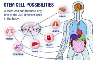
Recently in Ireland, people are paying thousands of euro for procedures advertised as ” stem cell treatment ”, for conditions such as osteoarthritis. These treatments are not covered by health insurance companies etc. They often use the term ”stem cell treatment”, for a process that bears little if any resemblance to what stem cell therapy actually is. Below is a summary of an article written by an orthopedic surgeon outlining the limitations of stem cell therapy as it presently stands. Before you part with your money for one of these treatments, have a read of it, discuss it with your doctor and do your research.


Below is a summary of an article written by Mark Miller, Professor of Orthopedic Surgery, University of Virginia, ”Stem cell treatment for arthritic knees is unproven, expensive and potentially dangerous”

Stem cells are “uncommitted” cells that are, at least theoretically, capable of becoming any type of cell – skin, heart, kidney or even knee cartilage cells. Stem cells can come from fetal tissue, including products of in-vitro fertilization as well as placenta and umbilical cord tissue. They can also come from a patient’s own “hidden” adult stem cells, which are most often harvested from bone marrow and fat. The potential for using these cells in medicine is tremendous.

The truth

Unfortunately, the excitement about stem cells has outpaced the science. In addition, due to the ethical issues associated with the use of fetal tissue, the U.S. Food and Drug Administration has severely restricted its use and obtaining an abundant source of concentrated stem cells can be difficult.

In orthopedics, researchers have proposed using stem cells for the treatment of joint/cartilage damage. This includes osteoarthritis. Osteoarthritis often results in the need for joint replacement surgery. Stem cell injections are now being  promoted as a potential way of avoiding the need joint replacement, by ”regenerating” the cartilage. Unfortunately, current technology and regulatory issues make obtaining and concentrating true stem cells a challenge, and encouraging them to become and remain cartilage cells and nothing else is even more difficult.

The problem with stem cells is that these cells can continue to evolve; they may not stop development at the cartilage cell phase. They may continue to differentiate into bone cells. This would make the joint even worse because bone creates a rough surface adjacent to the smooth articular cartilage. Bone is actually the end result of arthritis.

According to the American Association of Hip and Knee Surgeons, there are no proven medications or therapies that can delay or reverse the progressive joint destruction that occurs with osteoarthritis.

Many patients have paid out thousands of dollars for ”so-called stem cell treatment” only to later discover that they were scammed. Any positive effects of current stem cell treatment are likely not the result of the actual cells themselves but something else.

Alternatives to stem cell treatment

Depending on the cause and severity of their joint pain, patients have treatment options that range from physical therapy, to injections of various medications, to surgery. All have pros and cons; steroid injections can provide quick but short-lived pain relief, while a knee replacement can provide a permanent solution but also requires months of rehabilitation. Doctors need to help patients make the choice that best fits their particular needs.


Physiotherapists Tralee phone 086-7700191


Gout ?

Gout
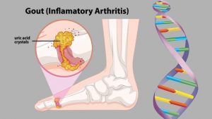
Gout is a form of arthritis which starts as a result of excess uric acid build-up in the blood, also called hyperuricemia. It is an extremely painful condition. Uric acid is produced in the body during the breakdown of purines – chemical compounds that are found in certain foods such as meat, poultry, and seafood. Normally, uric acid is dissolved in the blood and is excreted from the body in the urine via the kidneys. If too much uric acid is produced, it can build up and form needle-like crystals that trigger inflammation and pain in the joints and surrounding tissue. The condition often starts by affecting the joint at the base of the big toe. Attacks often occur without warning and in the middle of the night. They can come on quickly and keep returning over time, slowly damaging tissues in the region of the inflammation.

Tests and diagnosis

Gout can be tricky to diagnose, as it’s symptoms can be similar to those of other conditions, i.e. bunion on the big toe. While hyperuricemia occurs in the majority of people that develop gout, it may not be present during a flare. There is one very subtle, important difference between gout and other foot conditions, that helps the health practitioner differentiate between them. When you very gently stroke the skin of a gout affected area, it will cause a lot of pain in the sufferer. This does not occur with other conditions. The gout makes the skin super sensitive. This test is not 100% accurate, but I have found it to be a reasonably good indicator that one should look in the direction of gout, and refer the patient onto a doctor. This is especially true, if during examination and history take, the patient meets other criteria that would make them more prone to gout, and there has been no specific injury to the area affected. Often with tests you are more trying to rule out other conditions and narrow your possibilities down to that of gout..

One diagnostic test that doctors can carry out is the joint fluid test, where fluid is extracted from the affected joint with a needle. The fluid is then examined to see if any urate crystals are present. As joint infections can also cause similar symptoms to gout, a doctor can also look for bacteria when carrying out a joint fluid test in order to rule out a bacterial cause. Doctors can also do a blood test to measure the levels of uric acid in the blood. However, people with high uric acid levels do not always experience gout. Equally, some people can develop the symptoms without having increased levels of uric acid in the blood. Finally, doctors can search for urate crystals around joints or within a tophus(deposit of uric acid crystals) using ultrasound or CT scans.  X-rays cannot detect gout, but may be used to rule out other causes.

Treatment

Treatment and control of the condition requires certain dietary modifications such as reducing the intake of foods high in purines ie. red meat, shellfish, and beer to name but a few. Other factors that contribute to gout include being overweight, certain medications, high blood pressure, drinking too many sugary sodas, and being regularly dehydrated. These all need to be monitored and corrected.

Treatments for acute attacks include:

  1. NSAIDs – Non-Steroidal Anti-Inflammatory Drugs
  2. Colchicine
  3. Steroids
  4. Urate-Lowering Therapy (ULT)


For more see video


Overview by physiotherapists in Tralee, Co. Kerry


 Hammer Toes

Hammer toes and mallet toes are foot deformities that occur due to an imbalance in the muscles, tendons or ligaments that normally hold the toe straight. This is a deformity that causes your toe to bend or curl downward instead of pointing forward. It can affect any toe.

Hammer ToesHammer Toes

Causes of Hammer Toes

Hammertoe and mallet toe have been linked to:

  • An imbalance in toe muscles leading to instability, which in turn can cause the toe to contract.
  • Genetics(say one of your parents has it). It may be present at birth.
  • It may develop over time due to arthritis. Diabetes could also increase your risk.
  • An injury in which you stub, jam or break a toe can make it more likely for that digit to develop hammertoe.
  • Poorly fitting and/or high-heeled shoes or footwear that are too tight at the toes crowding your toes into a space in which they can’t lie flat.
  • toe length(if your second toe is too long)
  • Sex (women seem more prone but maybe that is down to fashion ie. high heels etc.?)

Complications of Hammer Toes

At first, a hammertoe or mallet toe might maintain its flexibility. Eventually, the tendons of the toe can contract and tighten, causing your toe to become permanently bent. The toe may become painful and moving it may also be difficult or painful. The raised portion of the toe or toes can rub against your shoes, causing painful corns or calluses.

Prevention of Hammer Toes

Relieving the pain and pressure of hammertoe may involve changing your footwear and/or wearing orthotics. If you have a more severe case of hammertoe or mallet toe, you might need surgery to get relief.

You can avoid many foot, heel and ankle problems with shoes that fit properly. Here’s what to look for when buying shoes:

  • Make sure there is adequate toe room. Avoid shoes with pointed toes.
  • Low heels. Avoiding high heels.
  • Make sure shoes are supportive and comfortable. Lace them up properly

These additional tips can help you buy the right shoes:

  • Buy shoes at the end of the day. Your feet swell slightly throughout the day.
  • Check your size. As you age, your shoe size might change — especially the width. Measure both feet and buy for the larger foot.
  • Buy shoes that fit. Be sure shoes are comfortable before you buy them. If necessary, a shoe repair store might be able to stretch shoes in tight spots, but it’s better to buy them to fit.

For more on hammer toes  see this video

Physiotherapy clinics in Tralee and Dingle phone Eddie on 086-7700191

Phones, laptops, reading in bed and the consequences of poor posture.

Poor Posture
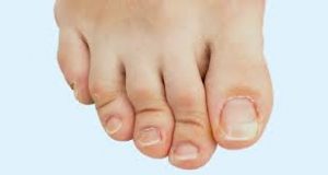
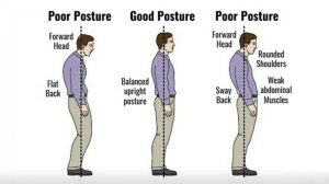
The increasing time we are spending glued to our phones or in front of a laptop with poor posture is manifesting itself with a large increase in the incidence of neck and shoulder pain presenting at clinic. Even kids are suffering from it. Good posture, as it relates to the neck, and outlined by a physiotherapist is commonly considered to be when the ears are positioned directly above the shoulders with the chest open and shoulders back. This neutral position minimizes the stress on the neck(cervical spine). Also it is important that the head is turned to full rotation left and right regularly in order to keep joints and muscles mobile and supple.

Imbalances from poor posture

Forward head posture occurs when the neck slants forward, placing the head further in front of the shoulders rather than directly above. This can lead to problems such as


  • Increased stress on the cervical spine. As the head moves forward in poor posture, the cervical spine must support an increased amount of weight.
  • Muscle overload. Forward head posture means muscles of the neck, shoulders and upper back have to continually overwork to counterbalance the pull of gravity of the forward head. As a result, muscles become more susceptible to painful strains and spasms.
  • Hunched upper back. Forward head posture is often accompanied by forward shoulders and a rounded upper back, which can increase the risk of developing pain and dysfunction in the neck, upper back, and/or shoulders.

The above can lead to more severe manifestations if the forward head posture is maintained long term. These include

  • Muscle imbalances. Some muscles in the neck, upper back, shoulders, and chest can become shortened and tight, whereas others can become elongated and weak.
  • Elevated risk for spinal degeneration. Extra stress on; the discs of the cervical spine’s, the facet joints, and vertebrae increases the risk of degenerative spinal issues, such as cervical degenerative disc disease and cervical osteoarthritis. It can also contribute to the development of bone spurs.
  • There is an increased risk of development of shoulder bursitis, specifically subacromial bursitis
  • Reduced mobility. With increased stiffness in the muscles and/or joints, the neck’s range of motion becomes decreased.
  • Nerve pain. Spinal degeneration and muscle spasm in the neck can lead to nerve compression resulting in neurogenic pain. This pain can be severely debilitating.

Treatment

Physiotherapist treatment of the above may consist of deep tissue massage, mobilizations and posture correction exercises. Severe cases of disc degeneration and/or nerve compression may require surgery. Mostly people suffer from the effects of poor posture later in life. The effects take time to manifest. Often people will only get the message of how detrimental poor posture can be when they start suffering from neuropraxia (nerve compression/ pain,tingling, numbness running down yours arms, worse at night). This condition can be quiet challenging for a physiotherapist treat. Physiotherapy treatment can still be very successful if it is caught in the early stages.


Physiotherapist in Tralee.

Iliotibial band syndrome. The I.T. band is not the problem.

iliotibial band syndrome

Regularly I come across  people receiving seemingly endless treatment sessions iliotibial band syndrome, with the main focus of the treatment being to try and loosen out the I.T. band. Below are a few things I believe you should consider when  treating  iliotibial band syndrome.

1. A client is told they have certain weak muscle groups which are causing the problem and are given a load of exercises to correct these weaknesses. Sometimes they are told that they need orthotics. Sometimes these treatment protocols may be necessary, but more often this is just an overuse injury that is being made way too complicated. Say you have been running for a year(or years) with no issues, why are all these ”weaknesses” just now causing you problems? Most likely they are not the cause.  Why do you suddenly need orthotics. Did you change your feet(joke)? It is more likely you are overdoing it a bit in your running.

2. All the emphasis is often placed on the I.T. band and treatment of same. The I.T. band is a long tendon and is a fairly inflexible structure, so concentrating on loosening out that is not going to give much relief in itself. Now if you shift the focus to loosening out the tightened outer quad muscles which attach to the I.T. band by fascia(connective, supportive sheet of tissue, kind of like a very strong clingfilm) and as a result pull on the I.T. band, you will generally start to recover fairly quickly. There is nearly always a section of the outer quad about a quarter of the way up from the knee which will be tight and ropy in this injury. Also don’t forget the Tensor fascia lata muscle. This is up near the hip. It is the muscle that attaches directly to the I.T. band. If this has tightened it also needs to be worked out and loosened also. The I.T band should not be totally ignored of course. Instrument assisted soft tissue release can be excellent in loosening it somewhat, as well as for loosening out tightened quad muscles and fascia.

3. Another problem with this condition is a lack of understanding as to what is causing the pain at the side of the knee. The tension and resulting friction on the I.T. band causes the bursa under it at the side of the knee to become inflamed. This is what causes the pain. Treatment options here include a combination of non steroidal anti-inflammatories, compression with ice(very effective), maybe some electro-acupuncture. You want to compress the icepack into the side of the knee to flatten the inflamed bursa and reduce inflammation. A packet of peas or one of those gel packs will not be cold enough.

4. Rest is needed. This is an overuse injury, remember, but the better and more effective the treatment, the faster the recovery.

Above is not necessarily stuff you will find in a text book but comes from knowledge of anatomy and experience in  successfully treating this condition hundreds of times over the years, often in about two treatment sessions. Injuries are often over-dramatized. Sometimes you just need a little rest, with the right physio treatment protocol to aid recovery.

Lumbar Spinal Stenosis

spinal stenosis
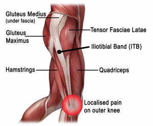
Spinal stenosis is a narrowing of the space within the spine. This can put pressure on the nerves that travel through the spine. It occurs most often in the lower back and the neck. Some people are born with a congenital form, but most develop it as part of the degenerative cascade. Sometimes people do not feel effects of the narrowing, but as part of the aging process, most people will eventually notice radiating pain, weakness, and/or numbness secondary to the compression of the nerves or spinal cord. In lumbar stenosis, the spinal nerve roots in the lower back become compressed and this can produce – tingling, weakness or numbness that radiates from the low back and into the buttocks and legs, especially with activity.

Lumbar spinal stenosis often mimics the symptoms of vascular insufficiency. Both conditions can cause claudication, which means leg pain with walking. If vascular studies identify normal blood flow, and tests confirm spinal stenosis,the symptoms are then referred to as neurogenic claudication.


Trigger Finger

trigger finger
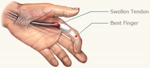
Trigger finger is a condition in which one of your fingers gets stuck in a bent position. Your finger may bend or straighten with a snap — like a trigger being pulled and released.

Tendons are fibrous cords that attach muscle to bone. Each tendon is surrounded by a protective sheath. Trigger finger occurs when the affected finger’s tendon sheath becomes irritated and inflamed. This interferes with the normal gliding motion of the tendon through the sheath. Prolonged irritation of the tendon sheath can produce scarring, thickening and the formation of bumps (nodules) in the tendon that impede the tendon’s motion even more.

People with work or hobbies that require repetitive gripping actions are at higher risk of developing trigger finger. The condition is also more common in women and people who suffer with diabetes.

Symptoms

Signs and symptoms of trigger finger may progress from mild to severe and include:

  • Finger stiffness, particularly in the morning
  • A popping or clicking sensation as you move your finger
  • Tenderness or a bump (nodule) on palmside at the base of the affected finger
  • Finger catching or locking in a bent position, which suddenly pops straight
  • Finger locked in a bent position, which you are unable to straighten

Trigger finger can affect any finger and triggering is usually more pronounced in the morning.


Diagnosis

Diagnosis of trigger finger doesn’t require any elaborate testing. Diagnosis is based on  medical history and a physical exam. During the physical exam, your doctor/physo will ask you to open and close your hand, checking for areas of pain, smoothness of motion and evidence of locking.  He should also feel your palm to see if there is a lump present. If the lump is associated with trigger finger, the lump will move as the finger moves because the lump is an area of swelling in part of the tendon that moves the finger.

Treatment

Trigger finger treatment varies depending on the severity and duration of the condition. Nonsteroidal anti-inflammatory drugs  may relieve the pain but are unlikely to relieve the swelling constricting the tendon sheath or trapping the tendon.

Conservative non-invasive treatments may include:

  • Rest. Avoid activities that require repetitive gripping, repeated grasping or the prolonged use of vibrating hand-held machinery until your symptoms improve. If you can’t avoid these activities altogether, padded gloves may offer some protection.
  • A splint. Your doctor may have you wear a splint at night to keep the affected finger in an extended position for up to six weeks. The splint helps rest the tendon.
  • Stretching exercises. Your doctor may also suggest gentle exercises to help maintain mobility in your finger.

If conservative treatment fails here are the other options.

  • Steroid injection. Injection of a steroid medication near to or into the tendon sheath may reduce inflammation and allow the tendon to glide freely again. This is the most common treatment, and it’s usually effective for a year or more in most people treated. Sometimes it takes more than one injection. For people with diabetes, steroid injections tend to be less effective.
  • Percutaneous release. After numbing your palm/finger, your doctor inserts a sturdy needle into the tissue around your affected tendon. Moving the needle and your finger helps break apart the constriction that is blocking the smooth motion of the tendon.This treatment may be done under ultrasound control, so the doctor can see where the tip of the needle is under the skin and to be sure it opens the tendon sheath without damaging the tendon or nearby nerves.
  • Surgery. Working through a small incision near the base of your affected finger, a surgeon can cut open the constricted section of tendon sheath. This is a last resort.


Update

Here is a video of exercises to get rid of trigger finger, I came across by the internet famous physical therapists Bob and Brad. These exercises have actually proved to be extremely effective  …….See video


Physio in Tralee phone 086-7700191