myofascial-pain-syndromeMyofascial Pain Syndrome

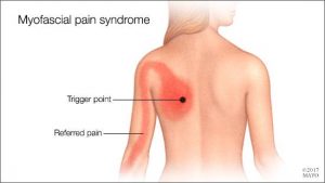
Myofascial pain syndrome is where pressure on sensitive points within your muscles (trigger points) causes pain and sometimes refers pain to seemingly unrelated parts of your body. It can occur after a muscle has been contracted repetitively ie. repetitive motions used in jobs or hobbies or by stress-related muscle tension. While nearly everyone has experienced muscle tension pain at some point, the discomfort associated with myofascial pain syndrome persists or worsens. There is no laboratory, radiographic or other diagnostic tests to prove the diagnosis of myofascial pain syndrome so it is considered a “subjective” diagnosis. Myofascial trigger points do not always cause pain. Sometimes they can lie dormant or inactive within a muscle for months or even years. Trigger points can often be identified by a skilled therapist working through your muscles with deep tissue massage. They feel like little knots deep within the muscles which when pressed are extremely tender.

Fascia is the body’s connective tissue. It is a head to toe, all-encompassing and interwoven system of fibrous connective tissue found throughout the body. Your  fascia provides a framework that helps support and protect individual muscle groups, organs, and the entire body as a unit. It is the same as that cling film, elastic type structure that surrounds a joint of meat, helping hold it together when the outer skin is removed.

This fascia in itself can also contribute to ”myofascial pain” syndrome. Injury, illness, stress, aging and repetitive use, can cause the fascia to shorten, thicken and become more unyielding . All the nerves and blood vessels run through the fascia. Therefore, if this connective tissue is tight, the associated tissues will have poor nutrient exchange. This exacerbates any painful situation because toxic metabolic waste products build up which often further aggravate pain receptors. This can create a vicious cycle, leading to increased muscle tension and further thickening and hardening of the fascia, which in turn further limits mobility.

 Symptoms

Signs and symptoms of myofascial pain syndrome may include:

  • Deep, aching pain in a muscle
  • Pain that persists or worsens
  • A tender knot in a muscle
  • Difficulty sleeping due to pain
  • Spasm in the area

The most commonly affected muscle groups include those of the neck, shoulders, upper  & lower back. Generally one side of the body is more affected than the other. It is common for patients with myofascial pain syndrome to have poor sleep patterns. This is associated with feeling unrested after a nights ”sleep” and daytime tiredness . Stiffness after inactivity is also a common feature.


Treatment

Myofascial pain can often benefit greatly from ”proper skilled deep tissue massage”, and trigger point release, both manual and with dry needling techniques. The fascia can also be stretched and worked out during the massage.  This is also one of the benefits of a stretching program when incorporated into your exercise routine. It helps keep prevent the fascia from tightening up because you are continually stretching it out.

During treatment for myofascial pain it is important that the patients reduce their stress levels, if this is a contributing factor to the condition. Exercises prescribed by a physio may also help, along with improving ones sleep patterns. In severe, chronic cases, medications may be needed to aid recovery.

Often trials of different medications are used to find the best treatment for a particular patient. For example, trazodone or amitriptyline may be used at bedtime to improve sleep as well as relieve pain;  cyclobenzaprine or  orphenadrine can also be  used to relax muscles and aid sleep; and antidepressants such as sertraline, fluoxetine(prozac), duloxetine, can be used to control pain, as can lyrica and gabapentin. Medications have side effects, so are  added as a last resort in chronic cases showing little improvement with physio alone. They should only be taken under a doctors supervision.


Physiotherapists Tralee : Phone 0867700191

Arthritis – Rheumatoid vs Osteoarthritis


Rheumatoid arthritis

rheumatoid arthritis
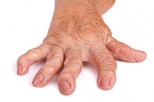
Rheumatoid arthritis is a chronic inflammatory disorder that can affect more than just your joints. It is an autoimmune disease occurring when your immune system mistakenly attacks the  synovium , the lining of the membranes that surround your joints. The resulting inflammation causes the synovium to thicken, eventually destroying the cartilage and bone within the joint. The tendons and ligaments holding the joint together also weaken and stretch. Gradually, the joint loses its shape and alignment.

It is unknown exactly  what starts the process, although a genetic component appears likely.  Newly developed  medications have greatly improved treatment options.  However, severe rheumatoid arthritis can still cause physical disabilities.

 Symptoms

Signs and symptoms of rheumatoid arthritis may include:

  • Tender, warm, swollen joints
  • Joint stiffness that is usually worse in the mornings and after inactivity
  • Fatigue, fever and weight loss

Early rheumatoid arthritis tends to affect your smaller joints first — particularly the joints of the fingers and toes. As the disease progresses, symptoms often spread to the wrists, knees, ankles, elbows, hips and shoulders. In most cases, symptoms occur in the same joints on both sides of your body.

Rheumatoid arthritis can also affect many non joint structures. This is the case in about 40 per cent of sufferers. These include:

  • Skin
  • Eyes
  • Lungs
  • Heart
  • Kidneys
  • Salivary glands
  • Nerve tissue
  • Bone marrow
  • Blood vessels

Rheumatoid arthritis signs and symptoms can vary in severity and may come and go. Periods of increased disease activity, called flare ups, alternate with periods of relative remission — when the swelling and pain fade or disappear.

Risk factors

  • Your sex : Women are more prone than men .
  • Age : Can occur at any age, but most common between the ages of 40 and 60.
  • Family history : Increased risk if there is a family history of the disease.
  • Smoking : Cigarette smoking increases your risk of developing rheumatoid arthritis.
  • Obesity: People who are overweight or obese appear to be at somewhat higher risk .

Diagnosis and Blood tests

People with rheumatoid arthritis often have an elevated erythrocyte sedimentation rate (ESR, or sed rate) or C-reactive protein (CRP), which may indicate the presence of an inflammatory process in the body. Other common blood tests look for rheumatoid factor and anti-cyclic citrullinated peptide (anti-CCP) antibodies. Rheumatoid arthritis can be difficult to diagnose in its early stages because the early signs and symptoms mimic those of many other diseases. There is no one blood test or physical finding alone that confirms diagnosis.

Imaging tests

Your doctor may recommend X-rays to help track the progression of rheumatoid arthritis in your joints over time. MRI and ultrasound tests can help your doctor judge the severity of the disease in your body.

Treatment

There is no cure for rheumatoid arthritis. Recent studies indicate that remission of symptoms is more likely when treatment begins early using medications known as disease-modifying antirheumatic drugs (DMARDs).

The types of medications recommended by your doctor will depend on the severity of your symptoms and how long you’ve had the rheumatoid arthritis. Medications used include :

  • NSAIDs : Nonsteroidal anti-inflammatory drugs (NSAIDs) can relieve pain and reduce inflammation.
  • Steroids : Corticosteroid medications, such as prednisone, reduce inflammation and pain and slow joint damage.
  • Disease-modifying antirheumatic drugs (DMARDs) : These drugs can slow the progression of rheumatoid arthritis and save the joints and other tissues from permanent damage. Common DMARDs include methotrexate (Trexall, Otrexup, Rasuvo), leflunomide (Arava), hydroxychloroquine (Plaquenil) and sulfasalazine (Azulfidine).
  • Biologic agents : Also known as biologic response modifiers. These are a newer class of DMARDs(Disease-modifying antirheumatic drugs) .These drugs can target parts of the immune system that trigger inflammation that causes joint and tissue damage. Biologic DMARDs are usually most effective when paired with a nonbiologic DMARD, such as methotrexate.

All the above drugs have side effects which need to be monitored.

Surgery

Medications can slow joint damage due to rheumatoid arthritis. When the damage becomes excessive surgery may need to be considered to repair the damaged joints.

Osteoarthritis

Osteoarthritis is the most common form of arthritis. It occurs when the cartilage that cushions the ends of bones in your joints gradually deteriorates. Cartilage is a firm, slippery tissue that permits nearly frictionless joint motion. In osteoarthritis, the smooth surface of the cartilage becomes roughed and worn. Eventually, if the cartilage wears down completely, you may be left with bone rubbing on bone. The disorder most commonly affects joints of the hands, knees, hips and spine.

Osteoarthritis symptoms can usually be effectively managed, although the underlying process cannot be reversed. Staying active, maintaining a healthy weight and other treatments may slow progression of the disease and help improve pain and joint function. When joint pain and damage is severe, doctors may suggest joint replacement surgery.

 Symptoms

Osteoarthritis symptoms often develop slowly  over time. Signs and symptoms may include:

  • Pain. Your joint may hurt during or after movement.
  • Tenderness. Your joint may feel tender when you apply light pressure to it.
  • Stiffness. Joint stiffness may be most noticeable when you wake up in the morning or after a period of inactivity.
  • Loss of flexibility. You may not be able to move your joint through its full range of motion.
  • Grating sensation. You may hear or feel a grating sensation when you use the joint.


Risk factors

Factors that may increase your risk of osteoarthritis include:

  • Older age. The risk of osteoarthritis increases with age as joints wear over time.
  • Sex. Women are more likely to develop osteoarthritis, though it isn’t clear why.
  • Obesity. Carrying extra body weight contributes to osteoarthritis in several ways, and the more you weigh, the greater your risk. Increased weight puts added stress on weight-bearing joints, such as your hips and knees. In addition, fat tissue produces proteins that may cause harmful inflammation in and around your joints.
  • Joint injuries. Injuries, such as those that occur when playing sports or from an accident, may increase the risk of osteoarthritis.
  • Certain occupations. If your job includes tasks that place repetitive stress on a particular joint, that joint may eventually develop osteoarthritis.
  • Genetics. Some people inherit a tendency to develop osteoarthritis.
  • Bone deformities. Some people are born with malformed joints or defective cartilage, which can increase the risk of osteoarthritis.


Summary

Rheumatoid arthritis is an auto-immune response where your body mistakenly attacks its own joints. Osteoarthritis is more or less due solely to wear on a particular joint over time.


Aside

Sometimes MRI results or Xray results  state there is osteoarthritis present. With age most of us have some level of osteoarthritis due to wear on our joints. The important thing which your doctor will discuss with you is whether this level is normal for your age. A person can have mild osteoarthritis with no pain or symptoms and may have nothing to worry about.


Physiotherapist Tralee phone 086-7700191

Ankle Sprains

Most people go over on their ankle( ankle sprains) at some stage in their lives. Usually they go over outwards and it is the outside of the ankle that gets injured. The ankle is stabilized by muscles, tendons and ligaments. Usually when you go over, it is an instantaneous thing where the muscles have failed to switch on and stabilize and the strain falls on the ligaments to protect you. The muscles also get strained as they try to correct the situation mid accident firing up too late. There are a lot of  ligaments in the ankle so the stability is quiet strong. Luckily, usually the ligaments strain rather than rupture. If they rupture  the sound will almost be like the sound of a tree branch breaking. You most likely will hear it. Luckily, because there are so many ligaments, even if one or two rupture, you can usually rehab the ankle fully. Protection such as strapping/taping or the likes of an ASO ankle brace may be needed thereafter, if some ligaments are torn fully, and you are involved in a sport where rapid twisting and turning  are part of the game and the training.


Treatment

Treatment of ankle sprains is pretty straight forward. One important point is that often most of the pain is due to the swelling that occurs rapidly post injury, so it is important to limit this. Immobilize the foot straight away. Start on NSAIDs(non steroid anti-inflammatories) immediately. Ice immediately(wet towel around iced, around the foot for about 10 mins every half hour or so). There are cold compression therapy machines which are excellent for this kind of injury. Obviously they are not always readily available.  They probably are a worthwhile investment for competitive clubs as they are not overly expensive.

A few days after ankle sprains physio can be commenced. This often includes gentle mobilization of the joints of the foot, massage to ease of strained/spasmed muscles and remove some of the swelling through the lymph nodes etc. Different grades of damage require slight modifications in approach but the basic idea is to get rid of inflammation, get movement back in the joints of the foot and relax down the strained muscles. Home rehab given to the patient involves self mobilization of the foot along with progressive strengthening exercises. Usually the condition does not require too many physio sessions but rather a progressive strengthening program prescribed by the physio and followed by the patient. Sometimes if pain lingers, the fibular head may have dropped slightly during the injury(so it is misplaced slightly) and it may need to be gently mobilized back into place using something like ”Mulligan Technique”. The movement of the fibular head can have been tiny, but the shift can cause considerable pain to linger if not rectified.  A good physio will always have done this during treatment anyway.

ankle sprains
ASO Ankle Brace

Rehab progression will involve things like calf raises, heel to toes taps, proprioception exercises, standing on one foot, standing up on toes of one foot, hopping on one foot, walking , jogging, jumping in various directions. It is recommended that an ASO ankle brace be worn during training and playing for several months after the injury to prevent recurrence. Strengthening work and controlled  training is done without the brace to ensure continued strengthening of the ankle. The ankle will not weaken as a result of the ankle brace as the muscles still have to fire during play, but continued strengthening and a build up of controlled exercise intensity without the ankle brace should also take place for full recovery.

Physiotherapists in Tralee phone 086-7700191


Sub-acromial bursitis. Overview by Physio in Tralee.


sub-acromial bursitis 
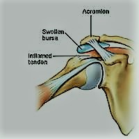
Sub-acromial bursitis is a common cause of shoulder pain that is usually related to impingement of the bursa between the supraspinatus muscle tendon and the acromion bone(see diagram). Bursae(single = bursa) are fluid-filled sacs that help reduce friction wherever tendons move under or over bone. The Supraspinatus muscle runs along the top of the shoulder blade and inserts via the tendon onto the top of the arm(humerus bone). This muscle is used to lift the arm up sideways . Above the supraspinatus tendon and under the acromion there is a bursa. When this bursa gets inflamed and swollen it can become trapped under the acromium bone of the shoulder causing pain and inflammation.


Symptoms

Symptoms of sub-acromial bursitis can be similar to those of supraspinatus  muscle/tendon injury within the shoulder. There will be pain and weakness in the arm, particularly when it is lifted sideways from the hip to overhead.  Pain at different levels 0f this 180 degree arc can indicate different injuries. If it is the tendon that is injured rather than the bursa somebody may be able to lift your arm over your head for you, with much less pain than you would have lifting your arm by itself. If you have a sub-acromial bursitis, especially if it is severe, neither you or another person will be able to lift that arm fully over your head. As the arm is lifted, there is increased compression on the bursa due to reduction within the sub-acromial space. This limits the upward movement of the arm  due to severe pain and restriction caused to the swollen/inflamed bursa. If a supraspinatus muscle/tendon tear is the cause of the pain, another person will be able to lift your arm fully over your head for you, with significantly less pain than you doing it on your own. This is  because they take over the function of the torn or injured muscle/tendon. These are important differences, as they often allow a practitioner to differentiate between both injuries .

What Causes It?

Sometimes, an injury damages the bursa in your shoulder. Overuse of your shoulder muscle can also cause damage. People who do a lot of overhead lifting and/or forceful pulling are at risk.  Sports  involving a lot of throwing or pitching can also irritate the sub-acromial bursa. Other factors that can help cause this type of bursitis include:

Your age. Bursitis in the shoulder becomes more likely as you age.

Poor posture with the shoulders arched forwards increases the risk of this injury. It causes impingement of the supraspinatus tendon and bursa by making the sub-acromial space smaller.

Poor shoulder flexibility/mobility.

Infection, arthritis, gout, diabetes, or thyroid disease can also cause issues.

Treatment

With very mild bursitis rest from aggravating activities and the use of non-steroid anti-inflammatories can be beneficial. A physio can loosen out the shoulder structure and give you exercises to improve shoulder posture, mobility and strength. In bad cases of bursitis, a cortisone injection into the area, done correctly, can bring full relief within days, especially if the condition is recent. Posture must be corrected, and aggravating activities reduced, thereafter, for long-term relief. For more troublesome recurring bursitis, a surgeon may need to remove the bursa altogether. Bursae do grow back, but now you have a new one to start afresh.

Physical Therapist in Tralee phone 086-7700191

Natural Anti-inflammatoriesNatural Anti-inflammatories

There are plenty advertisements for supplements like glucosamine and chondroitin claiming they will reduce inflammation and even rebuild cartilage etc. Most of these supplements have absolutely no evidence to back their claims, or they promote a few poorly carried out studies sponsored by the manufacturers. Three foods with quiet an amount of published scientific studies substantiating their usefulness as natural anti-inflammatories are turmeric, ginger and cinnamon.

Turmeric is a brilliant yellow spice commonly used in Indian cuisine and found in any grocery store. It is the curcumin in turmeric that has the anti-inflammatory properties.  You need a small amount of black pepper and fat in your food also to help with its absorption. Ginger is a zesty spice used in many cuisines. You can buy it powdered or as a fresh root in most supermarkets. Cinnamon is a popular spice often used to flavor baked treats. It is  better to use ceylon cinnamon rather than cassia cinnamon as the casia version is higher in toxins. These three spices have been used as medicines for centuries .  You can use all three(ginger, turmeric, cinnamon) in powder form, preferably organic.

Here is a little recipe that I got from the internet  and it actually  tastes quiet pleasant. Turmeric can be hard to eat unless it is hidden in a curry or a soup or something like that to mask the earthy flavor, but this recipe softens its taste.

1 cup of warm whole fat milk

1 teaspoon of turmeric powder

half teaspoon of ginger powder

half teaspoon of cinnamon powder

1 to 2 dessertspoons of honey

good pinch of black pepper

Blend it all up and drink. You would need to drink this daily to reduce inflammation.

Do the research online yourself and make up your own mind. Don’t use ”Google” as this fires everything at you both true and untrue. Key in ”Google Scholar”. This brings up a sub-site of Google which is more evidence based and shows all the scientific publications on your search.


Physiotherapy in Tralee – Phone 086-7700191

Tennis Elbow Misdiagnosis

Tennis Elbow Misdiagnosis

Tennis elbow, or lateral epicondylitis, is a painful condition of the elbow usually caused by overuse. Muscles, ligaments, and tendons support the functioning of the elbow joint. Your forearm extensor muscles and tendons extend the wrist and fingers. These extensor tendons attach the forearm muscles on the outside of the elbow to the lateral epicondyle. Tennis elbow is an injury to these tendons leading to pain and tenderness on the outside of the elbow. The main tendon involved in tennis elbow is usually the ”extensor carpi radialis brevis” tendon . Tennis elbow misdiagnosis is relatively common.

Tennis Elbow

When the tendon is weakened from overuse or trauma , microscopic tears form in the where the tendon attaches to the lateral epicondyle. This leads to pain and inflammation. The extensor carpi radialis brevis tendon may also be at increased risk of damage due to its position. As the elbow bends and straightens, the tendon rubs against the bone. This repetitive action can cause gradual wear and tear on the tendon over time in certain activities.

Activities – cause

Athletes are not the only people who get tennis elbow. Many people with tennis elbow participate in work or recreational activities that require repetitive and vigorous use of the forearm muscle. Painters, plumbers, carpenters, hairdressers, barbers, butchers(boners), people cutting hedges with clippers for long periods, are all particularly prone to developing tennis elbow.

Misdiagnosis

Tennis elbow misdiagnosis and mismanagement of this condition is often why people fail to recover. There are two common areas of misdiagnosis. Firstly if a person has not been doing any repetitive work with their forearm and there has been no forceful trauma while using the forearm, it is unlikely that the condition is tennis elbow. Sometimes the actual issue here is  nerve pain radiating down the arm from nerve compression in the neck and/or shoulder(neuropraxia). This nerve compression can be due to tight muscles/ locked facet joints and/or misaligned structures, often due to poor posture.  In the second type of tennis elbow misdiagnosis there can be repetitive injury involved but the condition is being exacerbated by nerve compression in the neck and shoulders, so both all areas need to be looked at for full resolution.

A cortisone injection is often used to try and treat ”tennis elbow”. This at most usually only gives short term relief and can even exacerbate the condition long-term with true tennis elbow, as it often allows the person to return to the aggravating activity thinking the injury has healed. Cortisone is an anti-inflammatory and may decrease inflammation in the area, reducing pain. It does not however stimulate tendon repair, which is what has been damaged in true ”tennis elbow” . Neither does cortisone do anything for a trapped nerve.

The above conditions are where an experienced physiotherapist/physical therapist can help greatly, usually much more so than any medication. This is their area of expertise.  However, you always need to remember accurate diagnosis and effective treatment is very practitioner dependent. Eccentric training along with soft tissue work can be very effective for true ”tennis elbow”. For the nerve compression (neuropraxia) described above, soft tissue release of neck and shoulder muscles(levator scapulae, trapezius,scalenes etc.) along with osteopathic manipulations to release any locked cervical or thoracic facet joints can be very effective. Symptoms of nerve entrapment/involvement can include neck/shoulder pain with tingling down the arms and/or into fingers. There are also manual orthopedic tests to check for nerve entrapment.

Aside : Carpal Tunnel syndrome is a medical condition due to compression of the median nerve as it travels through the wrist at the carpal tunnel. This can also be misdiagnosed and be due to nerve compression in neck /shoulder or upper thoracic regions.

Physiotherapists in Tralee  phone 086-7700191

IASTM
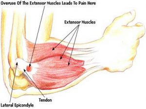
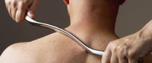
Instrument Assisted Soft Tissue Mobilization, also known as ”IASTM” for short is a process in which the clinician uses a set of ergonomically designed hand held instruments to break down the scar tissue and fascial restrictions in soft tissue (muscles, ligaments, tendons, and fascia ). The instruments you see in the photo are made of surgical grade stainless steel. Hypoallergenic aqueous cream or massage oil is used to facilitate gliding of the instrument along the muscle, tendon etc. during treatment.  The technique itself is said to have evolved from a form of Traditional Chinese Medicine called Gua Sha.  IASTM is a is a procedure that is growing  rapidly in popularity due to both the effectiveness and efficiency 0f the technique.

Once the damaged areas are detected, the instruments are used to deliver controlled microtrauma to the affected area. The purpose of the microtrauma is to stimulate a local inflammatory response, which initiates reabsorption of excessive scar tissue and facilitates a cascade of healing activities. Adhesions within the soft tissue that may have developed as a result of surgery, immobilization, muscle tears, or repetitive strain etc., are broken down, allowing the patient to regain function and range of movement. This treatment is a little intense, but it is extremely effective. It is somewhat similar to ”deep tissue massage” and/or ”rolfing”, but I always feel more is achieved in a shorter time-frame with IASTM technique . As in any Manual therapy treatment, it is usually not the only modality used. Supplementation with  stretching/strengthening exercises  etc. designed to correct biomechanical deficiencies by readdressing musculo-skeletal strength and imbalances  may also be prescribed  in conjunction with IASTM. I have used this form treatment both on myself and clients to great effect.

For more information check out these videos

Video 1

Video 2

Video 3


We are physiotherapists in Tralee, Co. Kerry. For more information on our treatments, prices, conditions we treat etc, check out our homepage. Also we are open 7am – 10pm weekdays and 8am – 2pm Saturdays. Phone 086-7700191


Muscle twitches

Muscle Twitches
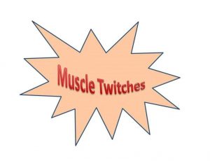
Muscles are made up of fibers that your nerves control. Muscle twitching is caused by minor muscle contraction in the area, or uncontrollable twitching of a muscle group that is served by a single motor nerve fiber. Stimulation or damage to a nerve may cause your muscle fibers to twitch. Most muscle twitches are minor and aren’t usually a cause for concern. Some are common and normal. Others are signs of a nervous system disorder.


Causes

These may include:

  • Autoimmune disorders such as Isaac syndrome.
  • Drug overdose (caffeine, amphetamines, or other stimulants).
  • Lack of sleep.
  • Drug side effect (such as from diuretics, corticosteroids, or estrogens).
  • Exercise (twitching is seen after exercise).
  • Lack of nutrients in the diet (deficiency).
  • Stress.
  • Medical conditions that cause metabolic disorders, including low potassium, and kidney disease, and uremia.
  • Twitches not caused by disease or disorders (benign twitches), often affecting the eyelids, calf, or thumb. These twitches are normal and quite common, and are often triggered by stress or anxiety . These twitches can come and go, and usually do not last for more than a few days.

Nervous system conditions that can cause muscle twitching include:

  • Amyotrophic lateral sclerosis (Lou Gehrig disease)
  • Neuropathy or damage to the nerve that leads to a muscle
  • Spinal muscular atrophy
  • Weak muscles (myopathy)


When to Contact a Medical Professional

Call your health care provider if you have long-term or persistent muscle twitches or if twitching occurs with weakness or loss of muscle.

Physiotherapist in Tralee : Phone 086-7700191

Arterial endofibrosis is an unusual condition, but an important one to keep in mind as a differential diagnosis, especially in the case of cyclists. When an athlete  bends forward and exercises(say like cyclists), it can put pressure on and/or cause a kink in the arteries supplying the working muscles of the leg with oxygen, sugars and so forth.  The artery affected  is usually one of the external iliac arteries which supply the majority of blood to each leg, from the heart.

Arterial Endofibrosis
Fig 1. Arterial Endofibrosis

The theory is that the longer time spent bending forward and exercising, the more the artery can be kinked and irritated. Over time, the body responds to this irritation by making the artery harder, thereby reducing the irritation to the artery (a good outcome), but this hardening also reduces the blood supply to the leg when exercising (a bad outcome for bike racers).

With Arterial endofibrosis the patient typically complains of weakness and cramping, ‘bursting’ or aching pain of a single leg only during heavy efforts on the bike.  They can’t ride through it, and in fact, the the harder they try, the worse it gets.  The onset is usually slow, over months, but extremely consistent. Always with increasing intensity comes decreasing strength.

The patient may get treated by a number of therapists often misdiagnosing the condition, and diagnosing some other exotic sounding condition instead. One thing remains constant though; the symptoms persist. A simple, but not always conclusive test that doctors perform is to exercise the patient, then take their blood pressure at their arm and at their ankle.  In a normal patient these two measurements will be the same or similar.  In a patient with the problem, the blood pressure in the ankle of the effected leg will be lower (because it is getting less blood).

More invasive tests may follow.   Treatment to correct the condition involves surgery. Often it is difficult to confirm the condition with absolute certainty.  This is important because the surgery carries risk, so you really want to be as sure as possible.  Surgery involves cutting out the kinked bit and sewing in another bit of vein from your leg instead, referred to as a ”patch”. Because a vein is a bit ‘flimsier’ than an artery, it bends a bit better. An artery has a muscular wall – which is why it pulses, making a patch using a piece of artery less suitable. Also you can ‘spare’ a bit of vein more than you can spare artery, and as a rule arteries run deep and are harder to harvest.

Return to cycling needs to be managed carefully post surgery, and has to be done under close medical supervision. However, the cyclist can eventually resume normal training/competition etc.

For more information see this video


Physiotherapists in Tralee. Open early until late. Phone 086-7700191.

Carpal tunnel syndrome
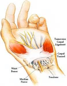
Carpal tunnel syndrome is a condition that causes numbness, tingling and weakness in the hand and arm. The condition is caused by  compression of the median nerve within the carpal tunnel, a narrow passageway on the palm side of your wrist. The median nerve runs from your neck  down along the arm and through the carpal tunnel to your hand. It provides sensation to the palm side of your thumb and fingers, except the little finger. It also provides nerve signals to move the muscles around the base of your thumb .  Anything that squeezes or irritates the median nerve in the carpal tunnel space may lead to this condition. The anatomy of your wrist, health problems and possibly repetitive hand motions may also contribute.   A wrist fracture can narrow the carpal tunnel and irritate the nerve, as can the swelling and inflammation resulting from rheumatoid arthritis.

Proper treatment usually relieves the tingling and numbness and restores wrist and hand function. It is worth noting that carpal tunnel syndrome is often misdiagnosed and the main site of median nerve compression can be occurring up at neck and shoulder level. Often by freeing up neck and shoulders muscles and surrounding joints with physio as well as working out muscles down along the arm through which the median nerve penetrates, and loosening and stretching the wrist structures, much relief, often total relief can often be achieved.


Symptoms

Syndrome symptoms usually start gradually.

Numbness: The first symptoms are usually tingling or numbness usually in the thumb, index and middle or ring fingers, but not your little finger. The sensation may travel from your wrist up your arm. These symptoms often occur while holding a steering wheel, phone or newspaper. The sensation may wake you from your sleep.

Weakness: You may experience weakness in your hand and a tendency to drop objects. This may be due to the numbness in your hand or weakness of the thumb’s pinching muscles, which are also controlled by the median nerve.


Carpal Tunnel Risk factors

A number of risk factors have been associated with carpal tunnel syndrome.  Although they may not directly be the cause , they may increase your chances of developing or aggravating median nerve damage.

These include:

Anatomic factors: wrist fracture, dislocation, or arthritis that deforms the small bones in the wrist, can alter the space within the carpal tunnel and put pressure on the median nerve.

Sex: It is generally more common in women. This may be because the carpal tunnel area is relatively smaller in women than in men.

Nerve-damaging conditions: Some chronic illnesses, such as diabetes, increase your risk of nerve damage.

Inflammatory conditions: Illnesses that are characterized by inflammation, such as rheumatoid arthritis, can affect the lining around the tendons in your wrist and put pressure on your median nerve.

Obesity : Being obese is a significant risk factor for carpal tunnel syndrome.
Alterations in the balance of body fluids. Fluid retention may increase the pressure within your carpal tunnel, irritating the median nerve. This is common during pregnancy and menopause. Carpal tunnel syndrome associated with pregnancy generally resolves on its own after pregnancy.


Carpal Tunnel Diagnosis

History of symptoms, Physical examination. Nerve tension tests.

X-ray. Some doctors recommend an X-ray of the affected wrist to exclude other causes of wrist pain, such as arthritis or a fracture.

Electromyogram. This test measures the tiny electrical discharges produced in muscles. During this test, your doctor inserts a thin-needle electrode into specific muscles to evaluate the electrical activity when muscles contract and rest. This test can identify muscle damage and also may rule out other conditions.

Nerve conduction study. In a variation of electromyography, two electrodes are taped to your skin. A small shock is passed through the median nerve to see if electrical impulses are slowed in the carpal tunnel. This test may be used to diagnose your condition and rule out other conditions.


Treatment

If the condition is diagnosed early, nonsurgical methods may help improve carpal tunnel syndrome. Physical therapy by a practitioner experienced in this area can be very effective. This would most like involve deep tissue work, osteopathic manipulations, electroacupuncture and postural correction exercises, and nerve glide exercises as the condition improves. Other treatment options include wrist splinting, taking more frequent breaks to rest your hands, avoiding activities that worsen symptoms, medications(Nonsteroidal anti-inflammatory drugs) icing, rest etc. Surgery is also an option but is a last resort.


For treatment of this condition contact us on 0867700191


For more information see this video