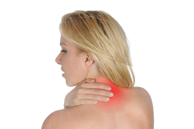
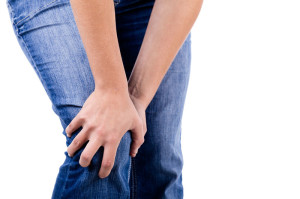
Muscle Tear or Spasm ? Which is it ?

muscle tearWhen somebody first presents in clinic with a muscle injury, it is important to first determine whether it is a muscle spasm or a tear. A spasm/cramp can often be worked out in a session with deep tissue massage and maybe some dry needling and stretching, allowing almost immediate turn to training. A muscle tear takes much longer to recover from, the length of time greatly depending on the degree of tearing. Treatment takes longer and a rehabilitation program is also essential.  A muscle spasm is caused by an involuntary contraction of that muscle. It is usually sudden, can cause significant pain and can limit the use of the muscle for a short period of time.

What is a Spasm?

 A muscle spasm is an involuntary contraction of that muscle. It is usually sudden, can cause significant pain and can limit the use of the muscle for a short period of time.

Spasm Causes

Spasms are usually caused by problems such as overworking a muscle, poor hydration, electrolyte imbalances, insufficient blood flow to the muscle or nerve compression. A pulled muscle is actually a tear in the muscle tissue. This occurs when the muscle is strained to the point where the muscle is damaged. The more significant the strain, the more the muscle can be torn.

What is a Muscle pull/Tear?

A pulled muscle is actually a tear in the muscle tissue. This occurs when the muscle is strained to the point where the muscle is damaged. The more significant the strain, the more the muscle can be torn.Pulled muscles are usually the result of not preparing the muscle for work through proper stretching, placing too much tension on the muscle or over-using the muscle. Soreness in the affected muscle is usually the first symptom. Using the muscle will be painful and difficult. In severe pulls, bruising may be present. The affected muscle will become very tender to the touch and some swelling may occur.

Pulled Muscle Causes

Pulled muscles are usually the result of one or a combination of (1) not preparing the muscle for work because of insufficient warmup,(2) lack of a proper stretching program after training to help the muscle recover,(3) placing too much tension on the muscle all of a sudden, (4) or over-using the muscle. Sharp pain in the affected muscle is usually the first symptom. Using the muscle will be painful and difficult. In severe pulls, bruising may be present. The affected muscle will become very tender to the touch and some swelling may occur.

The use of anabolic steroids is frequently linked to severe muscle tears as the body hasn’t had time to adapt to the increased workload . All training needs gradual progression and enough recovery time after training. The higher the intensity in training , the more time needed to recover. So for an athlete, periods of maximal performance require more recovery time.

For treatment of muscle tears or spasms phone 086-7700191. We are physiotherapists in Tralee.


Low Back Pain – Facet vs Disc

Two of the most  common causes of low back pain presenting in clinic are discogenic (disc) and facet joint related pain. Injury to either can cause severe discomfort, limiting a person’s ability to carry out normal daily activities. Facet joints are small articulations along each segment at the back of the spine, and help control the movement of the spine.  There are two of these joints at each vertebral level. There are intervertebral discs between each level  and these act as primary shock absorbers. They are  generally between a quarter and a half inch in height, interconnecting the bodies of the vertebral segments.  It can be considered that the discs and the facets create joint complexes that allow for both shock absorption and movement at each segment of the spine.

Low back pain
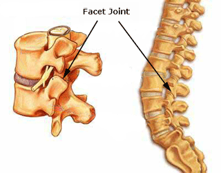
One of the primary symptoms when a  lumbar facet joint locks is an increase in pain on extension of the lower back.  Basically there is more pain when you try and straighten up your lower back or lean back on it. This position loads pressure on the locked lumbar facet joints.   Usually with this type of injury there is pain relief to a certain extent when a person bends forward.  The reason for this is that forward flexion decompresses the facet joint articulations, releasing some of the pressure on the joints and hence the associated pain. There can be nerve irritation with this injury but it is usually more general and not as severe as with discogenic pain.

Low back pain
Disc disorders

When we start talking about disc injury, we are usually referring to a small tear in the outer annular fibers of a disc that has either resulted in a bulge, a protrusion, or an extrusion of the disc particle(see diagram).  Pain occurs when a disc touches off a nerve due to one of these injuries, triggering a series of reactions, some of which include muscle spasm, inflammation and pain.   Often the nerve root irritation sends pain signals down either the front or back of the leg depending on which disc level is affected. In contrast to facet joint injuries, with disc injuries, bending forward causes a significant increase in pain, as it usually increases compression of the disc against the nerve.  So again to generalize, disc injuries are usually more sensitive to forward flexion,  whereby facet injuries are very sensitive to backward extension. A classic and very telling sign of a disc injury is where the spine becomes visibly curved off to one side as the disc tries to get away from the nerve it is touching. You will visibly see the person’s spine twisted off to one side.

With a locked facet joint, a simple osteopathic manipulation can give a person an immediate 60 – 70 % relief from pain symptoms.  The person is often almost back to normal the next day. With a facet joint injury, you are simply unlocking the locked facet joint in which the limited movement and was causing the problem. With a disc injury, you are trying to take pressure off the disc in the hope that it stops pressing against the nerve. Once off lumbar facet joint manipulation can play it’s part in recovery from disc disorders, if used at the right time by an experienced practitioner. Again this is used to mobilize the area and try and take pressure off the disc.

Often with a suspected disc injury a physio etc. may need to refer a patient to a doctor for a second opinion and most possibly medication( NSAIDs, muscle relaxant, nerve blocker), depending on the level of pain and discomfort. The doctor may also refer the patient for an MRI if they need more information on the level of damage. Physio generally involves deep tissue massage, dry needling, spinal manipulation at the right time. A rehab program involving pilates type exercises and some gentle stretching exercises etc. is paramount once a patient starts to improve. Continuous spinal manipulations several times a week for extended periods pushed by certain professions could do more harm than good, causing sheering of the disc and slowing healing.

”Putting back in a slipped disc” by manipulation is a myth. When somebody gets a manipulation done, the little crack you hear is when the facet joint gaps or unlocks. It is not the disc going back into place. Sometimes unlocking a facet joint in the area, if it has locked up say due to muscle spasm etc. can help take pressure of the disc in the area.

Eddie O Grady Physiotherapy, Tralee, Co. Kerry


Meniscus Injuries  – Knee Pain – Physiotherapy in Tralee

Cartilage within the knee joint provides cushioning between the bones at this joint. This protects them from the stresses of walking, jumping, running etc. There is articular cartilage which is the smooth, white tissue that covers the ends of bones(Femur, tibia) where they come together to form the knee joint. Healthy cartilage in our joints makes it easier to move. It allows the bones to glide over each other with very little friction. Articular meniscus injuries
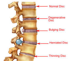
cartilage can be damaged by injury or normal wear and tear.

Within the knee you also have fibrocartilage in the form of the medial and lateral meniscii. These are two thick wedge-shaped pads of cartilage attached to top of the tibia (tibial plateau) and under the femur bone. They  allow the femur to glide when the knee joint moves. Each meniscus is curved in a C-shape, with the front part of the cartilage called the anterior horn and the back part called the posterior horn. Meniscal tears are usually described by where they are located anatomically in the C shape and by their appearance (for example, “bucket handle” tear, longitudinal, parrot beak, and transverse).

Blood supply

Because the blood supply is different to each part of the meniscus. Knowing where the tear is located may help decide how easily an injury might heal (with or without surgery). The better the blood supply, the better the potential for recovery. The outside rim of cartilage has better blood supply than the central part of the “C.” Blood supply to knee cartilage also decreases with age, with up to 20% of normal blood supply  lost by age 40.

What causes a meniscus to tear?

A forceful twist or sudden stop can cause the end of the femur to grind into the top of the tibia, pinching and potentially tearing the cartilage of the meniscus. These meniscus injuries can also occur with deep squatting or kneeling, especially when lifting a heavy weight. Meniscus tear injuries often occur during athletic activities, especially in contact sports like football and hockey. Motions that require pivoting and sudden stops, in sports like tennis, basketball, and golf, can also cause meniscus damage.

Increasing risk with age

The risk of developing a torn meniscus increases with age. This is  because cartilage begins to gradually wear out, losing its blood supply and its resilience. Being overweight also puts more stress on the meniscii. This means that routine daily activities like walking and climbing stairs increase the potential for wear, degeneration, and tearing. It is estimated that six out of 10 patients older than 65 years have a degenerative meniscus tear. Many of these tears may never cause problems.

Some of the fibers of the cartilage are interconnected with those of the ligaments that surround the knee. Thus, meniscus injuries may also be associated with tears of the collateral and cruciate ligaments, depending upon the mechanism of injury.

Symptoms of a meniscus injury can include some or all of the following:

  • Pain with running or walking longer distances
  • Intermittent swelling of the knee joint: Many times, the knee with a torn meniscus feels “tight.”
  • Popping, especially when climbing up or down stairs
  • Giving way or buckling (the sensation that the knee is unstable and the feeling that the knee will give way): Less commonly, the knee actually will give way and cause the patient to fall.
  • Locking (a mechanical block where the knee cannot be fully extended or straightened): This occurs when a piece of torn meniscus folds on itself and blocks full range of motion of the knee joint. The knee gets “stuck,” usually flexed between 15 and 30 degrees and cannot bend or straighten from that position.  

Diagnosis

The diagnosis of a knee injury begins with a history of the injury etc. and physical examination. There have been many tests described to assess the internal structures of the knee. The McMurray test is one long used orthopedic test . The health-care professional flexes the knee and rotates the tibia while feeling along the joint. The test is positive for a potential tear if a click is felt or noticeable pain is felt while circumducting the knee in full flexion.

Physiotherapy in Tralee – referral for MRI

(MRI) is the test of choice to confirm the diagnosis of a torn meniscus. It also allows a radiographer to visualize the inner structures of the knee. These structure include; the cartilage and ligaments, the surface of the bones, and the muscles and tendons that surround the knee joint. Plain X-rays cannot be used to identify meniscal tears but may be helpful in looking for bony changes, including fractures, arthritis, and loose bony fragments within the joint. In older patients, X-rays may be taken of both knees while the patient is standing. This allows the joint spaces to be compared to assess the degree of cartilage wear. Cartilage takes up space within the joint and if the joint space is narrowed, it may be an indicator that there is less cartilage present, likely from degenerative disease.

Treatment of Meniscal Tears

Sometimes conservative measures such as physical therapy, NSAIDs and rest can be enough to settle the condition. When conservative measures are ineffective the next step may be surgery to repair or remove the damaged cartilage.


For physiotherapy in Tralee phone 086-7700191, click here for website homepage

Levator Scupula Muscle Related Neck pain

When you wake up in the morning with neck pain, a crick in your neck, feel a burning pain on the top inner corner of your shoulder blade, or have trouble turning your head to look behind you while driving etc., the culprit may be a Levator Scapula muscle in spasm. The pain can be described as a throbbing, ache, or tightness. It usually presents from the top inner corner of the shoulder blade up along the neck. The Levator Scapulae muscles are located on either side of the neck. They originate on the four upper vertebrae of the cervical spine (neck). They insert, or attach, to the scapula, also known as the shoulder blade at the superior, medial border. These two muscles are involved in elevation, downward rotation and abduction of the scapulae. They are also involved in flexion and extension of the cervical spine(neck), turning of the neck slightly left and right, along with side bending of the neck left and right.


Neck pain
Levator Scapula Muscle Trigger points (Xs)
Neck pain
Levator Scapula Muscle

What Causes Levator Scapula Spasm / Trigger Points?

A trigger point is a tight area within muscle tissue that causes pain in that area and/or other parts of the body. The trigger points are shown above as two dark red circles, with the pain referral area also shaded in. Muscle spasm is a tightening of a muscle usually, due to overuse or overstress. It  can in itself cause pain and loss of mobility.

Causes

The following events and activities are likely to activate, or reactivate, tension / pain and trigger points in the levator scapulae.

  • whiplash from an automobile accident
  • sleeping on the stomach with the head turned/or sleeping in an odd position
  • chilling of the muscle during sleep from an air conditioner or draft from an open window
  • working at a computer with the head turned for long periods
  • holding a phone between the shoulder and ear
  • carrying a heavy bag with a shoulder strap
  • use of crutches that are too tall and elevate the shoulder
  • emotional and mental stress
  • Working with your arms raised above your head for prolonged periods of time can also irritate the Levator Scapula. To help reduce neck pain, stabilize your shoulder blade when you raise your arm.
  • poor posture with a forward head position puts this muscle under continuous strain causing overuse.

Treatment for Levator Scapula Muscle Spasm / Trigger points

Usually the condition settles after a few days if it is only a once off. Proper hands-on deep tissue massage and dry needling, when used together, can be great to settle the condition either in the short term, or if the condition has become more chronic. Also for the long term, correcting posture, stretching and strengthening of the upper back(particularly middle / lower traps, serratus posterior, rhomboids etc.) and the neck muscles(specifically the posterior neck muscles) can help prevent the condition from returning.

Posture

Posture is key to a healthy neck and spine. Try and sit up straight on a comfortable supportive chair, when at work or at home. Allow your shoulders to relax by using the arm rests of the chair. When at a computer, pull the screen close and try to get the screen up at eye level (say using books underneath it). This will mean your head won’t have to be protruding forward. The same applies when driving a car. Pull the seat in close to the steering wheel(within reason). Try to position yourself so the shoulders are relaxed and the head isn’t jutting forward. Activities like reading in bed, playing computer games for hours etc. can really tighten up the levator muscles. They are a disaster for thoracic and cervical posture in the long term.

For more information click to see this video


Physiotherapist in Tralee Phone 086-7700191


Restless Legs Syndrome 

RESTLESS LEG SYNDROME
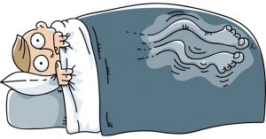
Restless legs syndrome(RLS) is a nervous system disorder resulting in an urge to move the legs. Because it usually interferes with sleep, it also is considered a sleep disorder. The condition causes an uncomfortable, “itchy,” “pins and needles,” or “creepy crawly” type feeling in the legs. These sensations are usually worse at rest, especially when lying or sitting. The symptoms are generally also worse in the evening and at night. The severity of RLS symptoms varies from mild to intolerable. Symptoms can come and go and vary widely in severity. For some people, symptoms may lead to severe sleep disturbance at night. This can significantly impair their quality of life.


Who Gets Restless Legs Syndrome?

It is estimated that ”Restless legs syndrome” (RLS) affects up to 10% of the population. It affects both sexes, but is more common in women. The condition may begin at any age, but most people affected are middle-aged or older. RLS is often unrecognized or misdiagnosed. This is especially true if the symptoms are intermittent or mild.


Causes of Restless Legs Syndrome

In most cases, doctors do not know the cause of restless legs syndrome. It is suspected that genetics plays a role. Nearly half of people with RLS also have a family member with the condition. Factors other than genetics are associated with the development of restless legs syndrome. Certain chronic diseases and medical conditions can cause RLS. These include iron deficiency, Parkinson’s disease, kidney failure, diabetes, and peripheral neuropathy. Treating these conditions often gives some relief from RLS symptoms.


Medications

Some types of medications may exacerbate symptoms. These include anti-nausea drugs, antipsychotic drugs, some antidepressants, cold and allergy medications containing sedating antihistamines. Women can experience RLS during pregnancy, especially in the last trimester. These symptoms usually go away within a month after delivery. Other factors, including alcohol use and sleep deprivation, may trigger symptoms or make them worse.


Diagnosis

There is no medical test to diagnose RLS. Doctors. However, blood tests or other diagnostic tests may be used to rule out other conditions. The diagnosis of RLS is based mainly on a patient’s symptoms and history .


Treatment

Treatment of RLS is targeted at easing the symptoms. In people with mild to moderate restless legs syndrome, lifestyle changes, such as beginning a regular exercise program , establishing regular sleep patterns, and eliminating or decreasing the use of caffeine,alcohol and tobacco, may be helpful.

Non-drug RLS treatments may include:

Leg massages

Hot baths or heating pads or ice packs applied to the legs

Good sleep habits

A vibrating pad called Relaxis


Drugs

Medications may sometimes be helpful in treatment of RLS but results vary among individuals. Drugs used to treat RLS include:

Dopaminergic drugs, which act on the neurotransmitter dopamine in the brain; Mirapex, Neupro, and Requip, levodopa.

Benzodiazepines, a class of sedative medications, may be used to help with sleep, but they can cause daytime drowsiness.

Narcotic pain relievers may be used for severe pain.

Anticonvulsant drugs  such as Tegretol, Lyrica, Neurontin,  and Horizant.


Although there is no cure for restless legs syndrome, current treatments can help control the condition, decrease symptoms, and improve sleep.


For more information see these videos     video 1.           video 2.


Restless legs syndrome treatment  by physical therapist in Tralee, Co. Kerry. Phone Eddie on 086-7700191. Click for Website

physiotherapist
Fig. 1. pronation-supination

Physiotherapist explains foot pronation & supination

A physiotherapist explains Pronation and supination as  movements that occur at the subtalar joint of the foot. The normal biomechanics of the foot are designed to absorb and direct the forces occurring throughout the gait cycle. As the foot is loaded, eversion of the subtalar joint, dorsiflextion of the ankle, and abduction of the forefoot occur. This is the pronation part of the gait cycle. Pronation should not continue past the latter stages of midstance during the gait cycle. At this stage the foot should then supinate in preparation for toe-off. Approximately four degrees of pronation and supination are necessary to enable the foot to propel forward properly. Any increase on this four degrees brings a foot into over-supination or over-pronation.


Pronation – physiotherapist explaination

Pronation of the foot is where the heel and the little toe move away from the center of the body. The foot also dorsiflexes up slightly, the ankle rolling inwards. Pronation is part of the natural movement of the human body. Certain injuries can occur with excessive pronation.  Runners with flat feet often tend to overpronate. Over-pronation can contribute to many injuries. These include shin splints, anterior compartment syndrome, patello-femoral pain syndrome, plantar fasciitis, tarsal tunnel syndrome, bunions (hallux valgus), achilles tendonopathies etc. The running shoes of over-pronators often show extra wear on the inner heel and ball of the foot.


Supination – physiotheraist explaination

With supination the heel and also the big toe rotate towards the centre of the body. The foot flexes down and the ankle rolls out. It is the opposite of pronation. A natural amount of supination occurs during the push-off phase of the running gait. This occurs as the heel lifts off the ground and the forefoot and toes are used to propel the body forward. However, excessive supination  places a large strain on the muscles and tendons that stabilize the ankle. This can make the oversupinator more prone to ankle sprain or ankle ligament rupture.

With over-supination the forces of impact on the foot are concentrated on a smaller area of the foot (the outside part), and are not distributed as efficiently. In the push-off phase, most of the work is done by the smaller toes on the outside of the foot, rather than the big toe. This places extra stress on the foot. It can lead to conditions such as iliotibial band syndrome, Achilles tendinitis, or plantar fasciitis. Over-supination causes the outer edge of running shoes to wear sooner. In extreme cases, there will also be holes in the uppers where the runner’s foot has broken through. Runners with high arches and tight Achilles tendons/calves tend to be over-supinators.


Orthotics

Properly prescribed foot orthotics can be beneficial in the treatment of over-pronation or over-supination. Physiotherapists usually supply both off the shelf and/or custom made orthotics. We  prescribe these only when we really feel they are necessary. Often the much cheaper off the shelf version will do the trick. It is important to get a quality product, prescribed by an experienced practitioner. Sometimes you just have an injury that needs  treatment and there is no need for an orthotic. Also high arches or a flat feet do not mean in itself you need orthotics, especially if you are not in pain.


Bone Spurs – Causes?

Bone spurs(also called osteophytes) are outgrowths of bone occurring along the edge of a bone. Bone spurs are most commonly found in joints, where two bones come together. They also occur close to where muscles, ligaments, or tendons attached to bone. Some common parts of the body affected include the neck (cervical spine), shoulders, hips, hands and heel bone.

Bone spurs typically occur because of continued stress or rubbing on a bone over a prolonged period of time. This can occur due to inflammatory conditions  such as osteoarthritis , tendinitis or tenosynovitis. Normally there is a smooth layer of cartilage on the edges of bones where they come together to form a joint. With osteoarthritis this cartilage layer becomes worn away. This causes the exposed bones to rub against each other. New bone forms in response to this stress and the resulting inflammation. Bone spurs usually develop in areas near tendons and ligaments due to chronic inflammation in these areas. This inflammation can result from friction between these tissues and bone, or from overuse. The bone spur development is the bodies way of trying to protect itself.

Signs and Symptoms

Bone Spurs
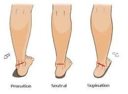
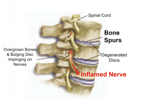
Bone spurs may or may not cause symptoms. Symptoms are location dependent. If bone spurs rub against other bones at joints they can cause pain and/or more limited movement in these joints. They can also be associated with pain, numbness, tenderness, and /or weakness in areas where  they irritate adjacent tissue structures(muscles, ligaments, bones, nerves etc.). If the bone spur rubs against tendons or ligaments, they can sometimes cause a tear in these tissues over time. This is a common cause of tears in certain rotator cuff tendons .

If bone spurs occur in the spine  they can sometimes pinch the nerves (radiculopathy) or spinal cord(myelopathy). A radiculopathy can cause pain, numbness, tingling, or weakness in the arms or legs depending on the area affected. With a myelopathy there can be pain and problems with balance along with weakness.

Diagnosis

Generally X-Rays, MRIs or CT scans are used to confirm suspected cases. Sometimes an experienced practitioner can feel the spur with their finger depending on the area(i.e. heel spur).

When Is Surgery Appropriate ?

In some cases, if symptoms cannot be controlled with more conservative treatment, surgery could be an option. The goal is to remove the bone spur, allowing a return to normal joint motion, or to remove the pressure on muscles, tendons, ligaments, or nerves.


Physiotherapist Tralee  :  Phone 086-7700191

”Shin Splints” – Which Type Have You ?

shin splints”Shin splints” is a catch-all term for shin pain either on the front outside part of the lower leg (anterior shin splints) or on the inside of the lower leg (medial shin splints). It is the curse of many athletes including runners, tennis players, dancers etc. Frequently the condition plagues novice runners who do not build their mileage gradually enough. It also affects seasoned runners who abruptly change their workout regimen, suddenly adding too much mileage, or switching from running on the flat to hills. The term mainly refers mainly to the following three conditions 1. mini stress fractures within the tibia bone, 2. chronic exertional compartment syndrome, 3. medial tibial stress syndrome.  It is important to differentiate between the three for treatment.


1. Real ”shin splints”

Real ”shin splints” are mini stress fractures (splint-ers) within the tibia bone. With this condition pain is gradual in onset, getting worse with activity, and there is usually a history of an increase in training intensity. Pain may occur with walking, at rest, or even at night in bed. Treatment for this condition involves rest for about eight weeks from running to allow the little stress fractures to heal. You should be able to keep up fitness levels by cycling, swimming etc., as these exercises are low impact. You may need to look at lower limb biomechanics, running style, training practices etc. to prevent recurrence of the shin splints .


2. Shin splints – Chronic exertional compartment syndrome

Chronic exertional compartment syndrome is defined as increased pressure within a closed fibro-osseous space(like the space the tibia and fibula), causing reduced blood flow and tissue perfusion(perfusion is the process of a body delivering blood to a capillary bed in its biological tissue), which subsequently leads to ischemic pain(pain due to restriction of blood supply, and thus oxygen and nutrients to tissue) and possible permanent damage to tissues of the compartment. The syndrome is frequently bilateral (both legs). Typical features of the condition are absence of pain at rest, with increasingly achy pain and a sensation of tightness in the shins upon exertion. Symptoms usually resolve or significantly dissipate within several minutes of resting. Anyone can develop the condition, but it is more common in athletes who participate in activities that involve repetitive impact, such as running. Sometimes Chronic exertional compartment syndrome may respond to deep tissue work and myofascial release of the structures involved. Changing your chosen activity to one involving less impact may also help. Surgery may be used as a last resort to relieve the pressure. It involves operating on the inelastic tissue encasing each muscle compartment (fascia). Methods include either cutting open the fascia of each affected compartment (fasciotomy) or actually removing part of the fascia (fasciectomy).


Shin splints – Medial tibial stress syndrome

 Medial tibial stress syndrome is an inflammation of the muscles, tendons, and bone tissue around your tibia. A common cause of Medial Tibial Stress Syndrome is pes planus (flat feet) or over-pronation of the foot during running. This puts increased strain on the Tibialis Posterior and soleus muscles leading to chronic traction at their insertions onto the periosteum on the posterior inner border of the tibia, producing pain in this area. Mild swelling in the area may also occur. The pain may be sharp and razor-like or dull and throbbing, occurring both during and after exercise, and aggravated by touching the sore spot. Initial treatment involves rest, ice, analgesics. Again switching to low impact activities such as swimming or cycling can keep a sports person active during recovery. For treatment, the entire calf should be assessed. The use of myofascial release techniques along with proper hands-on deep tissue work concentrating on thickened muscle fibres of the soleus, flexor digitorum longus and tibialis posterior adjacent to their bony attachments can prove effective. Dry needling and electro-acupuncture can also benefit recovery. Arch supporting orthotic insoles designed to reduce impact forces, correct flat-footedness and overpronation during running can help prevent recurrence and facilitate recovery by offloading affected structures. For some more information click here.

Dead leg

Dead Leg
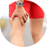
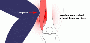
A ”dead leg”, also known as ”charley horse” or ”quadriceps contusion”, is essentially an injury due to a traumatic blow, crushing the quadriceps muscle against the femur bone. The quadriceps is the muscle at the front of your thigh. The injury can be either intermuscular or intramuscular. Treatment depends on the type of contusion and grade in severity of the injury. An Intramuscular contusion occurs when the muscle gets torn within the sheath surrounding it. This causes the initial bleeding to cease within hours due to increased pressure within the muscle. However, the fluid and blood is not able to escape from the muscle sheath surrounding it resulting in considerable loss of function and a lot of pain. This can take days or weeks for a full  recovery. You are unlikely to see any bruising with this type of contusion, especially in the early stages. In the case of intermuscular contusions, the muscle as well as part of the sheath surrounding it gets torn. This results in a longer bleeding time initially, especially if there is no use of ice therapy. The patient usually recovers faster from this type of dead leg, as the blood and fluids can easily flow away from the injury site. Bruising is often present in this type of contusion.

Grading

  • Grade 1: Quadriceps pain, tightness in the thigh and a limp while walking. The swelling is very mild and so is the pain. The patient has almost complete range of motion upon stretching.
  • Grade 2: The patient is not able to walk properly and limps when walking. There is some swelling present and the patient will experience bouts of pain upon activity. There is pain upon extending the leg against resistance. Pressure on the site also produces pain and the patient is unable to completely flex the knee.
  • Grade 3:The patient has severe pain with obvious swelling, and is unable to walk without the help of crutches. Muscle contraction produces an obvious gap or bulge. This type of dead leg needs at least a month or two to completely heal.

Treatment

  • I.C.E (Rest, Ice, Compression, Elevation).
  • Rest is very important and the limb should be kept elevated as much as possible.
  • Compression and ice therapy should be applied for 15 to 20 minutes every hour for the first 24 to 48 hours. Ice should be wrapped in a wet towel to prevent ice burns. A compression bandage should be worn 24/7 to reduce swelling and to give support until the pain has subsided.
  • Crutches should be used if required.
  • Never apply heat therapy(that includes having a hot bath…Do not) or perform deep tissue massage in the initial acute stage (24 to 48 hours) of a dead leg injury. This can aggravate the injury or lead to myositis ossificans later. Sports massage to help recovery should be started once the acute phase (24 to 48 hours) of the injury has passed, provided it’s not an intramuscular contusion(This may need more time to settle and sometimes requires medical intervention).
  • Gentle stretching of the area should be done after the acute stage (24 to 48 hours) has passed, but not before, provided it does not cause too much pain. The stretch should be held for 30 seconds and should be repeated 4 to 5 times in a day. With severe (grade 3) intermuscular contusion there should be no overpressure with passive stretching for 7-10 days.


When Can Exercises Be Started?

  • For dead leg of grade 1 contusion of quadriceps, strengthening exercises can be started as soon as the pain has subsided. Patient should start with mild exercises and should move on to difficult ones as the dead leg pain.
  • For dead leg of grade 2 contusions of quadriceps, strengthening exercises should be started after a week or so. Patient should start with gentle warm up exercises and finish with good stretching exercises. Strengthening exercises should be continued for several weeks. Strengthening exercises should be started more than a week later and for grade 3 contusions of the quadriceps.
  • Gentle exercises such as cycling or swimming are nice gentle strengthening exercises to start with.


Myositis Ossificans

The more severe a contusion, the greater the risk of development of Myositis Ossificans, especially with poor treatment and management. In this condition calcification occurs within the healing hematoma(swelling of clotted blood within the muscle). The healing hematoma forms bone within the layers of affected muscle. Symptoms include overnight and morning pain, as well as pain on muscle contraction. It may be possible to feel a hard bump or ‘woody’ lump within the affected muscle tissue.  Stiffness and loss of knee range of movement are also common.  Sometimes surgery is required to remove the calcified tissue.

Physiotherapists in Tralee

Early referral – Why it is so important – Physio in Tralee

PhysioYou have been getting treatment for back pain from your physio, chiropractor or osteopath, twice a week for the past four weeks. Each time you lie there for the first fifteen minutes with a hot pack while somebody else is being treated at the same time. The therapist pops in, has a brief chat, does a quick manipulation, reassures you of your improvement, then books you in for your next appointment. You leave wondering if you are really getting any better, but console yourself with the fact that the practitioner told you that you are.

Recently a client told me a story of where they endured a situation like the above for six months. Finally they decided to go to a doctor, who referred them for an MRI. The results showed a stress fracture to one of the lumbar vertebrae of the spine. In this case the treatments had been exacerbating the condition, and what was needed was rest and immobilisation.

Spinal Manipulations

Also manipulations, generally speaking, need to be done only once. They are used mainly to open a locked joint. A decent amount of soft tissue and myofascial work should be done by a physio beforehand to open up and relax the area. Otherwise the joint may revert to its locked position again shortly after the physio has manipulated it. Also as a general rule there should be a noticeable improvement in a clients condition from physio treatment to physio treatment. To illustrate the importance of early recognition and referral by your physio, let us look at a few more sinister conditions that present as back pain, requiring referral to a doctor or specialist.

Differential Diagnosis

Spondylitis; Ankylosing spondylitis is a condition where there is chronic inflammation of the spine and sacroiliac joints. This causes pain and stiffness in and around the spine, including the neck and back. Over time this condition can lead to a complete cementing together (fusion) of the vertebrae, a process referred to as ankylosis . Ankylosis causes loss of mobility of the spine.

Spondylolysis; A common cause of low back pain in adolescent athletes. It can be seen on X-ray and is a stress fracture in one of the bones (vertebrae) that make up the spinal column. It usually affects the fifth lumbar vertebra in the lower back, and less commonly the fourth. If the stress fracture weakens the bone too much the vertebra can start to shift out of place. This condition is called spondylolisthesis.

Spondylolysthesis; Spondylolisthesis is a condition whereby one of the vertebra of the spine slips forward or backward on the next vertebra. Spondylolisthesis can lead to deformity of the spine as well as a narrowing of the spinal canal (central spinal stenosis) and compression of the exiting nerve roots (foraminal stenosis). Spondylolisthesis is more common in the lower back but can also occur in thoracic and cervical spine.

Arthritis ; various types including spondylitis, reactive arthritis, osteoarthritis, juvenile onset spondyloarthritis, enteropathic arthritis, rheumatoid arthritis, polymyalgia rheumatica etc. can all present as back pain.

kidney stones ; can cause back pain

.Osteoporosis ; Osteoporosis means porous bones. It is a silent disease that usually goes undiagnosed until a bone fracture occurs. Bone is a living tissue that is constantly being turned over. Bones need normal sex hormones, calcium, vitamin D, adequate calories, proteins and weight bearing/strengthening exercise to keep them healthy. As we get older, more bone is lost than is replaced, but people with Osteoporosis lose more bone than people who do not have the disease. This causes bones to become more fragile and break or fracture more easily.

Various cancers ; pancreatic, liver cancers etc. can cause back pain.

ovarian cysts ; Ovarian cysts are fluid-filled sacs or pockets within or on the surface the female ovary. A large ovarian cyst can cause abdominal discomfort and a dull ache that radiates into the lower back and thighs.

Spinal stenosis ; This is a narrowing of spaces in the spine causing pressure on the spinal cord and nerves. About 75% of cases of spinal stenosis occur in the low back. In most cases, the narrowing of the spine associated with stenosis compresses a nerve root, which can cause pain down the leg.

physio in Tralee

Basically what I am saying in this article is that if your condition is not improving from physio session to physio session you may need to go back to your doctor for further investigation. Just keep it in mind. A good physio will probably have already referred you.