thoracic outlet syndrome
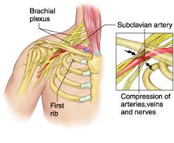
Thoracic outlet syndrome is a disorder that occurs when the blood vessels or nerves in the space between the collarbone and first rib (thoracic outlet) become compressed. This can result in any of the following symptoms ; 1. pain in your shoulders and/or neck , 2. numbness / pain / tingling down the arm and / or in a finger / fingers. Common causes include physical trauma, i.e. car crash ; repetitive injuries from job or sports related activities; anatomical defects (such as having an extra rib); poor posture( protruding neck / rounded shoulders); and pregnancy.

You may notice symptoms of thoracic outlet syndrome if your job requires you to do a repetitive movement continuously for long periods, i.e. typing on a computer; working on an assembly line; or lifting things above your head. These repetitive activities tend to tighten up neck and shoulder muscles. Athletes, such as baseball pitchers and swimmers can also develop thoracic outlet syndrome from the years of repetitive movements.

Obesity can put an undue amount of stress on your body structures and joints leading to the development of thoracic outlet syndrome. Carrying around an over sized bag or backpack or having somebody sitting on your shoulders for long periods, say at a concert can compress nerve structures etc. and lead to the condition. You may suffer symptoms during pregnancy as muscles, ligaments, tendons and joints come under strain. This is as a result of the release of the hormone relaxin around week twelve of pregnancy, and also due to the increase in bodyweight.

The two most common types of thoracic outlet syndrome are:

1. Neurogenic (neurological) : This form of thoracic outlet syndrome is characterized by compression of the brachial plexus ( nerves coming from the spinal cord (neck). This network of nerves controls muscle movements and sensations in the shoulder, arm and hand. This is the most common cause of thoracic outlet syndrome.

Signs and symptoms include:
• Numbness or tingling in your arm or fingers.
• Wasting of muscles through which the compressed nerve travels; either in the arm or fingers or both.
• Pain in the neck , shoulder or hand.
• Weakened grip in hand / loss of strength in the arm.

Treatment : Often if neurogenic thoracic outlet syndrome is caught early, deep tissue work on neck, shoulders and arm to loosen out tightened muscles; joint mobilisations / manipulation of the cervical and thoracic spine; electro acupuncture along the affected nerve pathway; postural correction exercises, can usually bring great relief within one or two treatment sessions. If the condition is chronic ( i.e. there a month or months), it may take more time for the irritated nerve to settle. You may need to lose weight if obesity is the cause of your thoracic outlet syndrome.

2.Vascular (venous or arterial): This type occurs when one or more of the veins or arteries are compressed in the area between the collarbone and first rib.
Signs and symptoms include:
• Discoloration of the hand (bluish colour).
• Arm pain and swelling, possibly due to blood clots.
• Blood clots in veins or arteries in the upper area of your body.
• Lack of colour in one or more of your fingers or your entire hand.
• Weakened or no pulse in the affected arm.
• Cold fingers, hands or arms.
• Arm fatigue after activity.
• Numbness or tingling in your fingers.
• Throbbing lump near your collar bone.
• Weakness of arm or neck.

Treatment : For vascular (venous or arterial) thoracic outlet syndrome…you need to consult with your doctor. This condition may require thrombolytic medications, anticoagulant medications or surgery.


Back Pain Treatment Options Made Simple

Three very common causes of back pain presenting in clinic are 1. muscular, 2. discogenic (from a disc), 3. locked facet joint. We will look at each and the relevant back pain treatment options.

Muscular back pain

For the low back a common muscle to cause trouble is the piriformis muscle (see diagram). It is involved in hip rotation movements so if it tightens you lose rotation putting pressure on the lower back during activities. When the piriformis muscle tightens it may also squeeze on the sciatic nerve which travels from the lumbar spine  down the back of the leg causing pain anywhere along the nerve path(low back, hamstring, calf, sole of foot). Tightened hamstrings may also contribute to low back pain(often resulting from being seated for long periods) making the spine do more work due to resulting poor posture in lifting etc.

The piriformis muscle can usually be worked out and loosened  with deep tissue massage.  The hamstrings can also be worked if these are contributing. Electro-acupuncture also helps loosen the muscles/trigger points and settle nerve irritation in affected areas.

Back pain

Locked Facet joint back pain

This is a common cause of back pain in the upper thoracic region(along back of the rib cage)…Often the person feels like somebody is sticking a finger into their back when they inhale deeply. Also with this condition a person may find that they can turn completely in one direction but not the other. In more serious cases pain may shoot down the arm due to nerve entrapment/irritation. A locked facet joint may also occur in the lower back sending nerve pain down the leg. The crack you hear when the back is manipulated is locked facet joints opening, it is not a ”slipped disc” being put back into place. A timely spinal manipulation done correctly, with a little soft tissue work done thereafter can often sort this condition. With the manipulation you get a little crack if the facet joint was locked, as it opens, giving immediate relief.

Back pain


Discogenic (Disc) related back pain

This is a more tricky one. Probably most common in the lower back (lumbar region). The patient will sometimes present with their spine all twisted off to one side in a sort of ”S” shape. This is usually because the disc bulge or protrusion is touching a nerve. The body compensates for this by trying to get the disc away from the nerve, hence the ”S” shape. Also there will be muscle spasm as the body tries to protect itself. A patient with this condition is usually in a lot of pain and even simple tasks like walking can be excruciatingly painful. Disc pain is less common in the thoracic region but again here it presents with deep unrelenting pain.

There is no such thing as a ”slipped disc”, it is just a misnomer. Discs basically bulge, degenerate(collapse) or herniate(a little fluid filled sack can protrude or leak out of them)(see diagram below). It is when this bulge or protrusion touches a nerve, the trouble starts.

Basically a muscle relaxant (valium) from your doctor along with an anti-inflammatory (NSAIDS) for a couple of days usually helps a lot.  Usually two treatment sessions along with the medication to loosen out the muscles will also reduce pain and improve recovery time. A manipulation to open any locked facet joints may help as the condition settles.

When a disc touches a nerve it initiates a series of reactions including muscle spasm and inflammation. The medication is very important in this condition and the combination of the both muscle relaxant and anti-inflammatory seem to complement each other along with treatment. Things such as ”Mckenzie technique” and ”nerve glides” can also help when introduced at the right time during treatment but require too much detail to go into here.

Once the condition settles it is important to 1. Train your body to engage its core. 2. Strengthen your core muscles with pilates type exercises, along with developing your core fitness. 3. Lose weight if you are overweight. 4. Try and improve your flexibity.

Once you get somebody moving, loosened out, give them a proper rehab routine and they are taking the medication, it is more about giving the body time to heal itself than anything else. Basically what you are trying to do with initial treatment is to relax any muscle spasm, reduce inflammation, reduce pressure on the disc with the more long-term goal of strengthening the body in the hope of bringing back in the disc bulge/ protusion enough from the nerve it is touching, so that symptoms subside. If there is little improvement in your condition after a week, an mri may be required to get more accurate information on the injury.  In some cases an epidural or surgery  may be required.

Back pain

Please note the above article only lists some common causes of back pain.

Back pain treatment in Kerry –  phone 086-7700191


Sever’s Disease – Kid’s Heel Pain

sever's disease
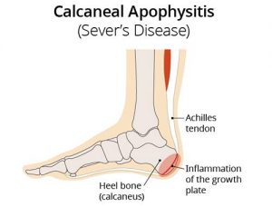
Sever’s disease (also known as calcaneal apophysitis) is a type of bone injury in kids whre the growth plate on the lower back part of the heel, where the Achilles tendon (the heel cord that attaches to the growth plate) attaches, becomes inflamed and painful. The reason for this is that the heel bone grows faster than the ligaments in the leg. As a result, muscles and tendons can become very tight and overstretched in children who are going through growth spurts. Sever’s disease occurs as a result of repetitive stress on the Achilles tendon. Over time, constant pressure on the already tight achilles tendon can damage the growth plate, causing children. Symptoms of Severs disease include pain and inflammation. This is why it is more common in physically active.

Symptoms of Sever’s disease include pain, inflammation or tenderness at the back of one or both heels which sometimes also extends in to the sides and bottom of the heel ; swelling and redness in the heel ; heel pain with limping, especially after running ; difficulty walking.Although it is painful and the name sounds a little scary, severs disease is only a temporary condition with no long-term problems. It is more common in physically active kids and occurs during the growth spurt at adolescence. This can begin any time between the ages of 8 and 13 for girls and 10 and 15 for boys. To be brief Sever’s disease is not a disease, it is in fact just growing pains.

Sever’s Disease – Treatment

The primary method of treating Sever’s disease is by taking time off or at least reducing participation in sports and other physical activities. This is in order to alleviate the pressure on the growth plate referred to above. Your physical therapist may prescribe stretching exercises for the calf muscles to help reduce pressure on the achille’s tendon attachment. Icing the area by placing an ice pack in a wet towel and applying it to the area of pain for 10 to 15 minutes at a time may help to alleviate pain and swelling, as may the use of NSAIDs(non steroidal anti-inflammatories). For more information on Sever’s disease click here.


Physiotherapists in Tralee, Co. Kerry : Phone 086-7700191

statinsStatins and Muscle Pain

Elevated low-density lipoprotein-cholesterol (LDL-C) has long been established as a major cause of coronary heart disease(CHD). The group of cholesterol-lowering drugs known as statins are widely used in the management of atherosclerotic disease processes that include CHD, myocardial infarction, stroke, and peripheral vascular disease(Pastemak et al. 2002). Although these drugs have been very successful in managing the cardiovascular health of many patients, they also can have some potential adverse side effects . These include muscle pain, inflammation, cramps and/ or weakness in areas such as the shoulder, neck, back, abdomen, hip, lower limbs, biceps brachii and masseter muscles etc.(Bennet et al. 2003). If left unchecked these symptoms may progress on to more serious conditions (Thompson et al. 2003) i.e. rhabdomyolysis. When detected early however, statin-related symptoms are reversible after withdrawal of the statin(Sinzinger et al. 2002). It is thus important for both physical therapists and physiotherapists to be aware of these associated symptoms linked to statin usage, because they frequently screen patients with musculoskeletal complaints. It becomes even more relevant when a patient presents with muscle pain, tenderness, weakness or inflammation that cannot be explained (for example, pain that is not due to physical work or injury).

Your doctor can carry out a blood test to measure a substance in your blood called creatine kinase (CK), which is released into the blood when your muscles are inflamed or damaged. If the level of CK in your blood is more than five times the normal level, your doctor may advise you to stop taking the statin. Regular exercise can also sometimes lead to a rise in CK, so tell your doctor if you have been exercising a lot. Once your CK levels have returned to normal, your doctor may suggest you start taking the statin again, but at a lower dose. It is important to note that a healthy diet and exercise can often reduce your LDL cholesterol to within the normal range without the need for medication. This is best done with the aid of a qualified dietician, backed up by follow up blood testing and advice from your doctor.

Statins and the problems found with grapefruit consumption

Grapefruit contains a chemical that interferes with your body’s ability to break down or metabolize certain statin medications. When statin takers ingest large amounts of grapefruit, the level of statins in their blood can increase, raising the possibility of side effects. Problems can occur for those who are sensitive to statin medications or those who have kidney disease or other illnesses. Side effects from grapefruit-statin interaction can be mild, such as muscle and joint pain. Severe side effects include muscle fiber breakdown and kidney failure.


References

Bennett W.E., Drake A.J., Shakir K.M. Reversible myopathy after statin therapy in patients with normal creatine kinase levels. Ann Intern Med.2003;138:436–437.

Pasternak R.C., Smith S.C., Bairey-Merz C.N., et al. ACC/AHA/NHLBI clinical advisory on the use and safety of statins. Circulation, 2002 ;106:1024–1028.

Sinzinger H., Wolfram R., Peskar B.A. Muscular side effects of statins. J Cardiovasc Pharmacol. 2002 ; 40:163–171.

Thompson P.D., Clarkson P., Karas R.H. Statin-associated myopathy. JAMA,2003 ;289:1681–1690.


Metatarsalgia

Metatarsalgia
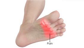
Metatarsalgia is the name given to pain in the front part of your foot under the heads of your metatarsal bones ( ball of foot, just before toes). It is usually worse when standing or walking etc.  and occurs most frequently in the second, third/or fourth metatarsal joints or isolated in the first metatarsal joint. Metatarsalgia usually comes on gradually over some weeks rather than suddenly. The affected area of your foot may also feel tender on palpation by your physiotherapist.

Common causes identified by physiotherapists include:

  • Pes cavus or high arched foot.
  • Excessive pronation of the foot(foot rools inwards ie. with flat foot).
  • Clawing or hammer toes.
  • Tight extensor tendons of the toes.
  • Prominent metatarsal heads.
  • Morton’s foot – here there is a shortened first metatarsal, which results in an abnormal gait putting increased pressure on the second metarsal.
  • Over doing it in athletes such as runners and tennis players etc. can lead to inflammation in the joints due to the pounding they receive.
  • Being overweight puts extra stress on many areas of the foot including metatarsals.
  • Wearing high heels – forward force on feet increases the pressure on metatarsal area.
  • Having tightened calf muscles or poor ankle flexibility – this affects the normal gait pattern.
  • Poorly fitting or tight footwear can squeeze the metatarsal joints causing friction , inflammation and poor gait.

Simple measures can help to relieve the symptoms of metatarsalgia. These include:

  • Resting with your feet elevated where possible.
  • Losing weight if you are overweight.
  • Wearing supportive shoes that are well fitted, low-heeled and have a wide toe area.
  • Metatarsal pads and orthotic inserts for your shoes may help to relieve pain in your forefoot by reducing the pressure placed on the heads of your metatarsal bones.
  • Physiotherapy may also be helpful. This may include stretching out the spaces between the metatarsal and mobilising the joints of the foot or performing deep tissue massage to loosen your calf muscles or any other lower limb muscles that may be contributing to poor gait. Sometimes if a metatarsal head has dropped it can be manipulated back into place. Pain relief as a result of this mobilisation, when effective can be immense and long lasting.
  • Simple painkillers such as paracetamol and non-steroidal anti-inflammatories(NSAIDs) such as ibuprofen may help to relieve pain.


Plantar Fasciitis – Foot Pain

plantar-fasciitis
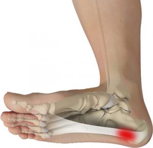
Plantar fasciitis is a painful inflammatory condition of the connective tissue on the sole of the foot(the plantar fascia). It is often caused by overuse of the plantar fascia, the tendons that help form the arch of the foot , running from the heel along the sole of the foot towards the toes. The plantar fascia  basically acts as shock absorber for the foot during movement while helping maintain the arch of the foot during standing. Plantar fasciitis is easiest treated when caught early. Longstanding cases often demonstrate more degenerative changes in the tissue than just inflammatory changes. This condition is called termedplantar fasciosis and can be much more difficult to treat.

While plantar fasciitis is often attributed to overuse in  athletes, among non-athletic populations it is associated with a high body mass index in combination with  long periods of weight bearing( Mc Poil et al. 2008).  Also people with flat feet or high arches tend to be more at risk.  A Flat foot tends to put increased strain on the origin of the plantar fascia at the heel (calcaneus) as the plantar fascia attempts to maintain a stable arch during the propulsive stage of the gait(walking, running etc.). Excessive movement of the forefoot in relation to the heel  during movement can also predispose to plantar fasciitis. In people with high arches there may be excessive strain on the heel due to the foots limited range of movement and thus a decreased ability to adapt  to the ground during movement.

Plantar fasciitis is commonly also associated with tightness in the calves, hamstrings and gluteal regions. Muscles in these areas have a significant effect on gait and thus foot biomechanics . Biomechanics is basically the science of movement, so when you here the phrase ”poor biomechanics” used it is basically referring to inefficiency during movement.

With the condition the pain is usually gradual in onset and felt on the medial aspect of the heel. Initially it is worse in the morning and decreases with activity, often aching afterwards. Periods of inactivity during the day are generally followed by an increase in pain as activity is recommenced. As the condition becomes more severe, the pain may be present when weight-bearing and worsen with activity.

Treatment

Effective treatment uses a multifaceted approach which may involve some or all of the following ; the use of nonsteroidal anti-inflammatory drugs (NSAIDs), deep tissue massage of the plantar fascia and along with deep tissue work to loosen out the calves, hamstrings and gluteal regions where necessary, stretching exercises for the plantar fascia(DiGiovanni  et al. 2003) and calf muscles, avoidance of aggravating activity, biomechanical correction with orthotics, electro-acupuncture and cold compression of the heel bursitis, strengthening exercises for the intrinsic muscles of the feet(Dyck and O’Neill ,2004), taping the heel into inversion (short term solution)( Radford  et al. 2006),  changing to proper supportive footwear containing well supported arches and midsoles (Yamashita, M.H. 2005). In extreme cases when all else has failed corticosteroid injections may be considered if there is a bursitis in the heel (Crawford et al. 1999) with surgery used as a last resort. This condition is usually resolved swiftly by an experienced practitioner without the need for surgery or corticosteroid injections.


Physiotherapist based in Tralee , Co. Kerry and open 7am to 11 pm weekdays, 7am to 2pm Saturdays.Please ring 086-7700191 anytime to make an appointment. We also specialize in proper deep tissue massage.


References

Crawford, E., Atkins, D., Young, P. et al. Steroid injection for heel pain: evidence of short term effectiveness: a randomized controlled trial. Rheumatology 1999;38(10):974-7.

DiGiovanni, B.F., Nawoczenski, D.A., Lintal, M.E. et al. Tissue-specific plantar fascia-stretching exercise enhances outcomes in patients with chronic heel pain. A prospective randomized study. J. Bone Joint Surg. Am. 2003;85-A(7):1270-7.

Dyck, D.D., Boyajian-O’Neill, L.A. Plantar fasciitis. Clin. Journal Sport Med. 2004;14:305-9.

McPoil, T.G., Martin, R.R.L., Cornwall, M.W. et al. Heel pain – plantar fasciitis: clinical practice guidelines linked to the international classification of functioning, disability, and health from the orthopedic section of the American Physical Therapy Association. J. Orthop. Sports Phys. Ther. 2008;38(4): A1-18.

Radford, J.A., Landorf, K.B., Buchbinder  R. et al. Effectiveness of low-dye taping foe the short-term treatment of plantar heel: a randomised trial. B.M.C.

Yamashita, M.H. Evaluation and selection of shoe wear and orthoses for the runner. Phys. Med. Rehabil. Clin. N. Am. 2005;16:801-29.