thoracic spine mobility
Thoracic Spine

A mobile thoracic spine allows you turn your back in many directions, enabling you to do everyday tasks with ease. Today’s sedentary lifestyle often contributes to reduced spinal mobility. Basically ”motion is lotion” and if you are inactive and also prone to poor posture, your thoracic spine can seize up. If it goes on long enough, say into old age portions of the spine may fuse and not move at all. A lack of thoracic spine mobility means that the lumbar spine, pelvis, shoulders and surrounding muscles  have to compensate. Long term, these over-compensations can  lead to overuse conditions and injuries, the lower back being particularly suceptable. This is because the lumbar spine is meant to keep us stable and is not very mobile, so when these joints are forced to overcompensate for the lack of movement in the thoracic area, it can place alot of pressure on the discs of the lower back. Possible consequences include inflammation, degeneration,  herniation of the discs, generalized low back pain, compression fractures, muscles spasms, and spinal nerve injuries. Similar pressures and injuries can occur in the neck and shoulders. For example, if your thoracic spine isn’t mobile, anytime you have to do a movement overhead, your shoulders make up for that lack of mobility. If you have shoulder impingement or chronic shoulder and neck problems lack of mobility in the thoracic spine will make every thing worse.


Improving Thoracic Spine Mobility

Yoga, pre- and post-workout stretching, and mobility exercises are the best way of maintaining and improving thoracic spine mobility. These need to be done regularly and consistently, especially as you get older. Your physiotherapist will be able to guide you on the correct exercises and help correct your form and technique until you get used to them and can do them yourself. Here are a few exercises to get you started.

For more information contact us @ Physiotherapists in Tralee.


Back Mice
Fig. 1 Back Mice

The term “back mice” is a rather cute description for a painful yet often overlooked condition, even by back pain specialists. The term  “back mice” was first used to label the condition by Peter Curtis in 1993. Back mice present as small, firm, fleshy yet moveable nodules upon palpation over the sacral region. Firm pressure directly on the nodules usually produces pain and tenderness, which sometimes radiates into the sacrum and hip. Also, the back mouse seems to suddenly appear following trauma to the back as in a motor vehicle accident or perhaps following a lifting injury. The size of the nodules does not change and they remain the same regardless of the administered soft tissue treatment.

Perhaps a more descriptive term for the back mouse is  “lumbar fascial fat herniation” . This occurs when the lumbar subfascial fat layer herniates through the overlying thoraco-dorsal fascia and gets trapped and inflamed. The mechanism appears to be due to an anatomical defect or weakened area in the fascia, which, when there is increased internal pressure, allows the fat lobules to push through the fascia. Once herniated, the fat becomes trapped and as an expanded, inflamed, herniation in an otherwise unyielding fibrous capsule. This creates a focus of pain. Pressure on the fat mouse does not push it back through the fascia but only inflames the torn fascia more. These herniations occur at predictable sites along the iliac crest and sacrum very close to the natural dimple area. They also are approximately three times more prevalent in women, particularly in moderately obese women.

A client usually presents with an episode of  low back pain. There may be a history of pain with lifting or prolonged sitting and the pain is usually greater on one side more than the other. The pain may radiate into the buttocks and sacrum and perhaps to the lateral thigh and into the lower extremity. Medications usually do nothing. Many will have gone through the pain pill merry-go-round, taking a cocktail of pain and anti-inflammatory medications even though the pain never truly goes away. The client has often tried everything, been everywhere and you are their last hope. They may have had radiographs, MRI studies or nerve conduction studies, all with negative or minimal findings. They may even have a minor disc bulge without nerve compression, yet the pain exactly mimics a discogenic disorder. When asked to point to the area of the greatest pain, they will invariably point just above and lateral to the natural “dimple” where the back and buttocks come together, near the multifidis triangle. When the area is palpated, the most marked finding is one or several firm, mobile 1.3 cm nodules. When pressed, these nodules reproduce the client’s complaints of back pain as well as the “sciatic” pain.

Subcutaneous lipomas in the back region differ from back mice in that they present as moveable non-tender “speed bumps” that cause pain only when they compress the underlying soft tissue. Subcutaneous lipomas can be found anywhere in the body. They grow slowly over time and are only cosmetically important.

back mice
Fig 2. Overlying thoraco-dorsal fascia.


back mice
Fig 3. Iliac crest and sacrum.


There always seems to be a focus by back pain specialists upon the disc and nerves issues even though the fat mice are readily palpable. Many sufferer’s have had epidural injections without success. I have had clients who have had surgery for disc herniations yet who still point to the back mouse post surgery as the focal point of pain. It could perhaps be considered diagnostic for the presence of a lumbar fascial fat herniation if a local infiltration of anesthetic takes away the pain.

Treatment of Back Mice

A medical doctor can inject the back mouse with a local anesthetic. This usually only helps temporarily. Dry needling techniques by acupuncturists may help reduce the tension in the fibrous capsule. Good results may sometimes be obtained with local electrical stimulation techniques such as electro-acupuncture. Do not apply deep pressure to back mice during manual physiotherapy treatment. Doing so may only serve to aggravate the herniation. Release of muscular tissue tension around the back mouse may provide some relief by easing pressure on the area.

Perhaps the only permanent cure for the back mouse is its excision and removal. This could be performed by a hernia repair specialist. Once the fat herniation is excised and the fascial tear repaired, the client usually enjoys a more enduring and sometimes dramatic relief. One of the biggest problems is that so many medical doctors fail to recognize this condition; they tend to discount its existence, thereby limiting the treatment options. Icing the area may also provide temporary relief.


For a short video on back mice click here.


Physiotherapist in Tralee. Phone 0867700191 to discuss your injury, make an appointment or get a second opinion.


stenosis of spineSpinal stenosis is a narrowing of the spaces within your spine, which can put pressure on the nerves that travel through the spine.  Others may experience pain, tingling, numbness and muscle weakness. Symptoms often start gradually and worsen over time. Spinal stenosis is most commonly caused by wear-and-tear changes in the spine related to osteoarthritis. In severe cases of spinal stenosis, doctors may recommend surgery to create additional space for the spinal cord or nerves. Most people with spinal stenosis are over the age of 50. There are two main types of spinal stenosis: 1. Cervical stenosis. where the narrowing occurs in  your neck. 2. Lumbar stenosis where the narrowing occurs in the lower back.


Symptoms of spinal stenosis

Neck (cervical spine)

  • Numbness or tingling in a hand, arm, foot or leg
  • Weakness in a hand, arm, foot or leg
  • Problems with walking and balance
  • Neck pain
  • In severe cases, bowel or bladder dysfunction (urinary urgency and incontinence)

Lower back (lumbar spine)

  • Numbness or tingling in a foot or leg
  • Weakness in a foot or leg
  • Pain or cramping in one or both legs when you stand for long periods of time or when you walk, which usually eases when you bend forward or sit
  • Back pain

Causes of spinal stenosis

The backbone (spine) runs from your neck to your lower back. The bones of your spine form a spinal canal, which protects your spinal cord (nerves). Some people are born with a small spinal canal. But most spinal stenosis occurs when something happens to narrow the open space within the spine, such as the following :

  • Overgrowth of bone. Wear and tear damage from osteoarthritis on your spinal bones can prompt the formation of bone spurs, which can grow into the spinal canal.
  • Herniated disks. The soft cushions that act as shock absorbers between your vertebrae tend to dry out with age. Cracks in a disk’s exterior may allow some of the soft inner material to escape and press on the spinal cord or nerves.
  • Thickened ligaments. The tough cords that help hold the bones of your spine together can become stiff and thickened over time. These thickened ligaments can bulge into the spinal canal.
  • Tumors. Abnormal growths can form inside the spinal cord, within the membranes that cover the spinal cord or in the space between the spinal cord and vertebrae. These are uncommon and identifiable on spine imaging with an MRI or CT.
  • Spinal injuries. Car accidents and other trauma can cause dislocations or fractures of one or more vertebrae. Displaced bone from a spinal fracture may damage the contents of the spinal canal. Swelling of nearby tissue immediately after back surgery also can put pressure on the spinal cord or nerves.


In severe cases of spinal stenosis, doctors may recommend surgery to create additional space for the spinal cord or nerves.


See video below for some tips.

https://youtu.be/GbJgE0LPKCk


Physio in Tralee specializing in the treatment of back pain, neck pain, foot pain and sports injuries.


When Back Pain Is A Symptom Rather Than A Condition

Back pain may sometimes be a symptom rather than a condition as such. A practitioner needs to always keep this at the back of their mind when treating or assessing somebody. Sometimes things need more investigation. A good physio will spot this early and refer you on quickly to your G.P. for further examination. It is always better to play it safe if in doubt.  I always believe you should be seeing improvement in your condition from treatment to treatment. If there is no improvement after several treatments you need to be asking yourself a few questions i.e. Do I need to try a different physio or do I need to consult my doctor for a second opinion. Sometimes the proper treatment protocol for a condition is all that is needed for a swift recovery. The list below is not meant to be alarmist. It is more to emphasize the fact that continuous unrelenting back pain can be a symptom of something else going on within your body.


Below are examples of conditions/medications etc. that may have back pain as a side effect/symptom.


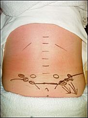
kidney stones – A kidney stone may not cause symptoms until it moves around within your kidney or passes into your ureter (the tube connecting the kidney and bladder). At that point, you may experience symptoms such as severe pain in your side and back below the ribs, or pain that radiates into the lower abdomen and groin.

Lupus is a long-term autoimmune disease in which the body’s immune system becomes hyperactive and attacks normal, healthy tissue. Symptoms include inflammation, swelling, and damage to the joints, skin, kidneys, blood, heart, and lungs. Lupus can cause neck and back pain, because muscles in these areas can become inflamed due to the lupus. Furthermore, the muscle pain syndrome  ”fibromyalgia”  can cause pain in these areas and is commonly associated with lupus.

Spinal arthritis/facet joint arthritis causes stiffness and back pain.

Cancers – A primary bone cancer tumor in the spine can cause back pain, as can a number of other cancers when they have metastasized(spread to other sites in the body), such as breast cancer, testicular cancer, colon cancer, and lung cancer. In fact, back pain is often the one of first symptoms that people with lung cancer notice before they are diagnosed. A tumor in the lungs can put pressure on the spine, or can affect the nerves around the chest wall and spine.

Spondylosis a painful condition of the spine resulting from the degeneration of the intervertebral discs.

Spondylitis is a condition resulting in inflammation within the joints of the spine. As the inflammation goes and healing takes place, bone grows out from both sides of the vertebrae and may join the two together, causing a stiffening known as ankylosis. The progressed condition is called ankylosing spondylitis. The cause is not yet known.

back pain


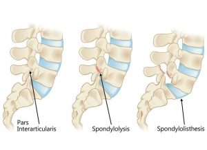
Spondylolisthesis is a slipping of vertebra that occurs, in most cases, at the base of the spine.

Spondylolysis is a defect or fracture of one or both wing-shaped parts of a vertebra, can result in vertebrae slipping backward, forward, or over a bone below.


Spondylosis, Spondylitis, Spondylolisthesis, Spondylolysis all have back pain as a symptom.


Fractures – Even a minor fracture along the spine  can cause considerable back pain.

StatinsStatins are drugs that can help lower your cholesterol. One of the more severe side effect of statins is myotoxicity(having a toxic effect on muscle), in the form of myopathy(a disease of the muscle in which the muscle fibers do not function properly. This results in muscular weakness), myalgia(muscle pain), myositis(inflammation and degeneration of muscle tissue) or rhabdomyolysis(a condition in which damaged skeletal muscle breaks down). Currently, the only effective treatment of statin-induced myopathy is the discontinuation of statin use in patients affected by muscle aches, pains and elevated creatine kinase levels. Creatine kinase are the clinical measure of muscle damage (rhabdomyolysis).

Anticonvulsant drugs such those used in the treatment of conditions like epilepsy can cause changes in calcium and bone metabolism. This may in time lead to decreased bone mass and a risk of osteoporotic fractures in the spine which may also result in severe back pain. Two widely used antiepileptic drugs phenytoin and carbamazepine are recognized to have direct effects on bone cells.

Corticosteroids – Longterm use of corticosteroids increase the risk of compression fractures in the spine(back).


Neurological Compromise — A Red Flag

Neural compromise can result from spinal cord or cauda equina compression . Cauda equina compression usually results from a fracture, tumor, epidural hematoma, or abscess, and occasionally from a massive disk herniation. Paraplegia(impairment in motor or sensory function of the lower extremities), quadriplegia(paralysis caused by illness or injury that results in the partial or total loss of use of all four limbs and torso), or cauda equina deficit should trigger an aggressive search for the cause.

Cauda equina compression classically presents with back pain, bilateral sciatica(pain down the back of both legs), saddle anesthesia, and lower extremity weakness progressing to paraplegia, but in practice these symptoms are variably present and diagnosing the condition often requires a high degree of suspicion. Hyporeflexia(no reflexes) is typically a sign of cauda equina compression, while hyperreflexia(overresponsive reflexes), clonus(series of involuntary, rhythmic, muscular contractions and relaxations), and the Babinski sign(see below *)  suggest spinal cord compression, requiring an evaluation of the cervical and thoracic spine. Cauda equina compression typically involves urinary retention; in contrast, cord compression typically causes incontinence. If either cauda equina or spinal cord compression is detected during an initial examination, an immediate more extensive evaluation is warranted. MRI is the study of choice.

*(The Babinski reflex occurs after the sole of the foot has been firmly stroked. The big toe then moves upward or toward the top surface of the foot. The other toes fan out. This reflex is normal in children up to 2 years old. It disappears as the child gets older. It may disappear as early as 12 months. When the Babinski reflex is present in a child older than 2 years or in an adult, it is often a sign of a central nervous system disorder. The central nervous system includes the brain and spinal cord.)

Spinal epidural hematoma

Spinal epidural hematoma is a rare but dramatic cause of paralysis in elderly patients. In most cases, there is no antecedent trauma. Lawton et al. (1995), in a series of 30 patients treated surgically for spinal epidural hematoma, found that 73% resulted from spine surgery, epidural catheterization, or anticoagulation therapy. Other possible causes of epidural hematoma include vascular malformations, angiomas, aneurysms, hypertension, and aspirin therapy. The same study found that the time from the first symptom to maximal neurologic deficit ranged from a few minutes to 4 days, with the average interval being nearly 13 hours.

Although painless onset has been reported, spinal epidural hematoma typically presents with acute pain at the level of the lesion, which is often rapidly followed by paraplegia(impairment in motor or sensory function of the lower extremities) or quadriplegia(is paralysis caused by illness or injury that results in the partial or total loss of use of all four limbs and torso), depending on the location of the hemorrhage. Sometimes the onset of pain is preceded by a sudden increase of venous pressure from coughing, sneezing, or straining at stool. Urinary retention often develops at an early stage.

Most lesions occur in the thoracic region(rib area of back) and extend into the cervicothoracic(upper back and neck) or the thoracolumbar(ribs to lower back area) area. The pain distribution may be radicular(affecting or relating to the root of a spinal nerve), mimicking a ruptured intervertebral disk.

Evaluation should be with MRI. Early recognition, MRI confirmation, and treatment should be accomplished as soon as possible. Recovery depends on the severity of the neurologic deficit and the duration of symptoms before treatment. Lawton et al.(1995), found that patients taken to surgery within 12 hours had better neurologic outcomes than patients with identical preoperative neurologic status whose surgery was delayed beyond 12 hours. Surgery should not be withheld because of advanced age or poor health: in 10 reported cases in which surgery was delayed, all patients died.


For more about treatments and opening times etc. click on our homepage
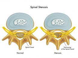
Everyone’s spine has curves, from your neck down to your lower back. These curves, which create the spine’s ”S” shape, are  lordotic in shape in the neck and lower back, and  kyphotic in shape for the upper back. They help your body 1. absorb shock,  2. support the weight of the head, 3. align your head over your pelvis, 4. stabilize and maintain its structure, 5. move and bend flexibly. When these curves are exaggerated through poor posture or hereditary conditions like scoliosis, spinal pain and problems can develop over time.


Kyphosis

Kyphosis
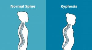
Kyphosis is an exaggerated, forward rounding of the back. It can occur at any age but is more common in older people. Age-related kyphosis is often due to weakness in the spinal bones that causes them to compress or crack. Other types of kyphosis can appear in infants or teens due to malformation of the spine or wedging of the spinal bones over time. Mild kyphosis causes few problems. Severe kyphosis can cause pain and be disfigurment. Treatment for kyphosis depends on your age, and both the cause and the effects of the curvature.


Lordosis

Lordosis
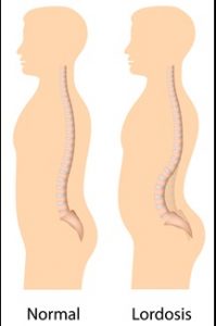
Some curvature in the lower back is normal. However, if your curve arches too far inward, it’s called lordosis, or swayback. Lordosis can affect your lower back and/or neck. The condition can lead to excess pressure on the spine, causing pain and discomfort. It can affect your ability to move if it’s severe and left untreated. Treatment of lordosis depends on how serious the curve is and how you got lordosis. There’s little medical concern if your lower back curve reverses itself when you bend forward. You can probably manage your condition with physical therapy and daily exercises.


Scoliosis

scoliosis
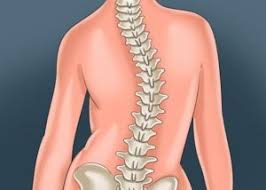
Scoliosis is a medical condition in which a person’s spine curves sideways, usually in an  “S” or “C” shape. In some, the degree of curvature  remains stable, while in others, it increases over time. Mild scoliosis does not typically cause problems, however severe cases can interfere with breathing. The cause of most cases is unknown, but is believed to involve a combination of genetic and environmental factors. Diagnosis is confirmed by Xray. Treatment depends on the degree of curve, location, and cause.

Minor curves may simply be watched periodically. Treatments may include bracing or surgery in more severe cases. The brace must be fitted to the person and used daily until growing stops. Evidence that chiropractic manipulation dietary supplements, or exercises can prevent the condition from worsening is non existent. However, exercise is still recommended due to its other health benefits. Scoliosis occurs in about 3% of people. It most commonly occurs between the ages of 10 and 20. Girls typically are more severely affected than boys.


Physiotherapists in Tralee Phone 086-7700191

Low Back Pain – Facet vs Disc

Two of the most  common causes of low back pain presenting in clinic are discogenic (disc) and facet joint related pain. Injury to either can cause severe discomfort, limiting a person’s ability to carry out normal daily activities. Facet joints are small articulations along each segment at the back of the spine, and help control the movement of the spine.  There are two of these joints at each vertebral level. There are intervertebral discs between each level  and these act as primary shock absorbers. They are  generally between a quarter and a half inch in height, interconnecting the bodies of the vertebral segments.  It can be considered that the discs and the facets create joint complexes that allow for both shock absorption and movement at each segment of the spine.

Low back pain
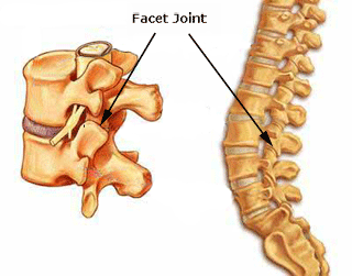
One of the primary symptoms when a  lumbar facet joint locks is an increase in pain on extension of the lower back.  Basically there is more pain when you try and straighten up your lower back or lean back on it. This position loads pressure on the locked lumbar facet joints.   Usually with this type of injury there is pain relief to a certain extent when a person bends forward.  The reason for this is that forward flexion decompresses the facet joint articulations, releasing some of the pressure on the joints and hence the associated pain. There can be nerve irritation with this injury but it is usually more general and not as severe as with discogenic pain.

Low back pain
Disc disorders

When we start talking about disc injury, we are usually referring to a small tear in the outer annular fibers of a disc that has either resulted in a bulge, a protrusion, or an extrusion of the disc particle(see diagram).  Pain occurs when a disc touches off a nerve due to one of these injuries, triggering a series of reactions, some of which include muscle spasm, inflammation and pain.   Often the nerve root irritation sends pain signals down either the front or back of the leg depending on which disc level is affected. In contrast to facet joint injuries, with disc injuries, bending forward causes a significant increase in pain, as it usually increases compression of the disc against the nerve.  So again to generalize, disc injuries are usually more sensitive to forward flexion,  whereby facet injuries are very sensitive to backward extension. A classic and very telling sign of a disc injury is where the spine becomes visibly curved off to one side as the disc tries to get away from the nerve it is touching. You will visibly see the person’s spine twisted off to one side.

With a locked facet joint, a simple osteopathic manipulation can give a person an immediate 60 – 70 % relief from pain symptoms.  The person is often almost back to normal the next day. With a facet joint injury, you are simply unlocking the locked facet joint in which the limited movement and was causing the problem. With a disc injury, you are trying to take pressure off the disc in the hope that it stops pressing against the nerve. Once off lumbar facet joint manipulation can play it’s part in recovery from disc disorders, if used at the right time by an experienced practitioner. Again this is used to mobilize the area and try and take pressure off the disc.

Often with a suspected disc injury a physio etc. may need to refer a patient to a doctor for a second opinion and most possibly medication( NSAIDs, muscle relaxant, nerve blocker), depending on the level of pain and discomfort. The doctor may also refer the patient for an MRI if they need more information on the level of damage. Physio generally involves deep tissue massage, dry needling, spinal manipulation at the right time. A rehab program involving pilates type exercises and some gentle stretching exercises etc. is paramount once a patient starts to improve. Continuous spinal manipulations several times a week for extended periods pushed by certain professions could do more harm than good, causing sheering of the disc and slowing healing.

”Putting back in a slipped disc” by manipulation is a myth. When somebody gets a manipulation done, the little crack you hear is when the facet joint gaps or unlocks. It is not the disc going back into place. Sometimes unlocking a facet joint in the area, if it has locked up say due to muscle spasm etc. can help take pressure of the disc in the area.

Eddie O Grady Physiotherapy, Tralee, Co. Kerry


Back Pain Treatment Options Made Simple

Three very common causes of back pain presenting in clinic are 1. muscular, 2. discogenic (from a disc), 3. locked facet joint. We will look at each and the relevant back pain treatment options.

Muscular back pain

For the low back a common muscle to cause trouble is the piriformis muscle (see diagram). It is involved in hip rotation movements so if it tightens you lose rotation putting pressure on the lower back during activities. When the piriformis muscle tightens it may also squeeze on the sciatic nerve which travels from the lumbar spine  down the back of the leg causing pain anywhere along the nerve path(low back, hamstring, calf, sole of foot). Tightened hamstrings may also contribute to low back pain(often resulting from being seated for long periods) making the spine do more work due to resulting poor posture in lifting etc.

The piriformis muscle can usually be worked out and loosened  with deep tissue massage.  The hamstrings can also be worked if these are contributing. Electro-acupuncture also helps loosen the muscles/trigger points and settle nerve irritation in affected areas.

Back pain

Locked Facet joint back pain

This is a common cause of back pain in the upper thoracic region(along back of the rib cage)…Often the person feels like somebody is sticking a finger into their back when they inhale deeply. Also with this condition a person may find that they can turn completely in one direction but not the other. In more serious cases pain may shoot down the arm due to nerve entrapment/irritation. A locked facet joint may also occur in the lower back sending nerve pain down the leg. The crack you hear when the back is manipulated is locked facet joints opening, it is not a ”slipped disc” being put back into place. A timely spinal manipulation done correctly, with a little soft tissue work done thereafter can often sort this condition. With the manipulation you get a little crack if the facet joint was locked, as it opens, giving immediate relief.

Back pain


Discogenic (Disc) related back pain

This is a more tricky one. Probably most common in the lower back (lumbar region). The patient will sometimes present with their spine all twisted off to one side in a sort of ”S” shape. This is usually because the disc bulge or protrusion is touching a nerve. The body compensates for this by trying to get the disc away from the nerve, hence the ”S” shape. Also there will be muscle spasm as the body tries to protect itself. A patient with this condition is usually in a lot of pain and even simple tasks like walking can be excruciatingly painful. Disc pain is less common in the thoracic region but again here it presents with deep unrelenting pain.

There is no such thing as a ”slipped disc”, it is just a misnomer. Discs basically bulge, degenerate(collapse) or herniate(a little fluid filled sack can protrude or leak out of them)(see diagram below). It is when this bulge or protrusion touches a nerve, the trouble starts.

Basically a muscle relaxant (valium) from your doctor along with an anti-inflammatory (NSAIDS) for a couple of days usually helps a lot.  Usually two treatment sessions along with the medication to loosen out the muscles will also reduce pain and improve recovery time. A manipulation to open any locked facet joints may help as the condition settles.

When a disc touches a nerve it initiates a series of reactions including muscle spasm and inflammation. The medication is very important in this condition and the combination of the both muscle relaxant and anti-inflammatory seem to complement each other along with treatment. Things such as ”Mckenzie technique” and ”nerve glides” can also help when introduced at the right time during treatment but require too much detail to go into here.

Once the condition settles it is important to 1. Train your body to engage its core. 2. Strengthen your core muscles with pilates type exercises, along with developing your core fitness. 3. Lose weight if you are overweight. 4. Try and improve your flexibity.

Once you get somebody moving, loosened out, give them a proper rehab routine and they are taking the medication, it is more about giving the body time to heal itself than anything else. Basically what you are trying to do with initial treatment is to relax any muscle spasm, reduce inflammation, reduce pressure on the disc with the more long-term goal of strengthening the body in the hope of bringing back in the disc bulge/ protusion enough from the nerve it is touching, so that symptoms subside. If there is little improvement in your condition after a week, an mri may be required to get more accurate information on the injury.  In some cases an epidural or surgery  may be required.

Back pain

Please note the above article only lists some common causes of back pain.

Back pain treatment in Kerry –  phone 086-7700191