Everyone’s spine has curves, from your neck down to your lower back. These curves, which create the spine’s ”S” shape, are  lordotic in shape in the neck and lower back, and  kyphotic in shape for the upper back. They help your body 1. absorb shock,  2. support the weight of the head, 3. align your head over your pelvis, 4. stabilize and maintain its structure, 5. move and bend flexibly. When these curves are exaggerated through poor posture or hereditary conditions like scoliosis, spinal pain and problems can develop over time.


Kyphosis

Kyphosis
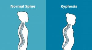
Kyphosis is an exaggerated, forward rounding of the back. It can occur at any age but is more common in older people. Age-related kyphosis is often due to weakness in the spinal bones that causes them to compress or crack. Other types of kyphosis can appear in infants or teens due to malformation of the spine or wedging of the spinal bones over time. Mild kyphosis causes few problems. Severe kyphosis can cause pain and be disfigurment. Treatment for kyphosis depends on your age, and both the cause and the effects of the curvature.


Lordosis

Lordosis
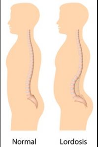
Some curvature in the lower back is normal. However, if your curve arches too far inward, it’s called lordosis, or swayback. Lordosis can affect your lower back and/or neck. The condition can lead to excess pressure on the spine, causing pain and discomfort. It can affect your ability to move if it’s severe and left untreated. Treatment of lordosis depends on how serious the curve is and how you got lordosis. There’s little medical concern if your lower back curve reverses itself when you bend forward. You can probably manage your condition with physical therapy and daily exercises.


Scoliosis

scoliosis
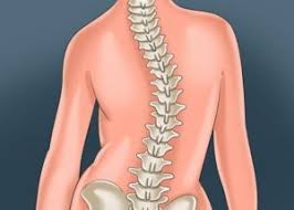
Scoliosis is a medical condition in which a person’s spine curves sideways, usually in an  “S” or “C” shape. In some, the degree of curvature  remains stable, while in others, it increases over time. Mild scoliosis does not typically cause problems, however severe cases can interfere with breathing. The cause of most cases is unknown, but is believed to involve a combination of genetic and environmental factors. Diagnosis is confirmed by Xray. Treatment depends on the degree of curve, location, and cause.

Minor curves may simply be watched periodically. Treatments may include bracing or surgery in more severe cases. The brace must be fitted to the person and used daily until growing stops. Evidence that chiropractic manipulation dietary supplements, or exercises can prevent the condition from worsening is non existent. However, exercise is still recommended due to its other health benefits. Scoliosis occurs in about 3% of people. It most commonly occurs between the ages of 10 and 20. Girls typically are more severely affected than boys.


Physiotherapists in Tralee Phone 086-7700191

Meniscus Injuries  – Knee Pain – Physiotherapy in Tralee

Cartilage within the knee joint provides cushioning between the bones at this joint. This protects them from the stresses of walking, jumping, running etc. There is articular cartilage which is the smooth, white tissue that covers the ends of bones(Femur, tibia) where they come together to form the knee joint. Healthy cartilage in our joints makes it easier to move. It allows the bones to glide over each other with very little friction. Articular meniscus injuries
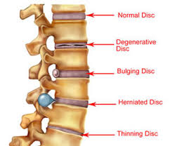
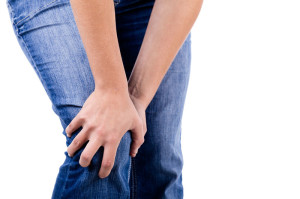
cartilage can be damaged by injury or normal wear and tear.

Within the knee you also have fibrocartilage in the form of the medial and lateral meniscii. These are two thick wedge-shaped pads of cartilage attached to top of the tibia (tibial plateau) and under the femur bone. They  allow the femur to glide when the knee joint moves. Each meniscus is curved in a C-shape, with the front part of the cartilage called the anterior horn and the back part called the posterior horn. Meniscal tears are usually described by where they are located anatomically in the C shape and by their appearance (for example, “bucket handle” tear, longitudinal, parrot beak, and transverse).

Blood supply

Because the blood supply is different to each part of the meniscus. Knowing where the tear is located may help decide how easily an injury might heal (with or without surgery). The better the blood supply, the better the potential for recovery. The outside rim of cartilage has better blood supply than the central part of the “C.” Blood supply to knee cartilage also decreases with age, with up to 20% of normal blood supply  lost by age 40.

What causes a meniscus to tear?

A forceful twist or sudden stop can cause the end of the femur to grind into the top of the tibia, pinching and potentially tearing the cartilage of the meniscus. These meniscus injuries can also occur with deep squatting or kneeling, especially when lifting a heavy weight. Meniscus tear injuries often occur during athletic activities, especially in contact sports like football and hockey. Motions that require pivoting and sudden stops, in sports like tennis, basketball, and golf, can also cause meniscus damage.

Increasing risk with age

The risk of developing a torn meniscus increases with age. This is  because cartilage begins to gradually wear out, losing its blood supply and its resilience. Being overweight also puts more stress on the meniscii. This means that routine daily activities like walking and climbing stairs increase the potential for wear, degeneration, and tearing. It is estimated that six out of 10 patients older than 65 years have a degenerative meniscus tear. Many of these tears may never cause problems.

Some of the fibers of the cartilage are interconnected with those of the ligaments that surround the knee. Thus, meniscus injuries may also be associated with tears of the collateral and cruciate ligaments, depending upon the mechanism of injury.

Symptoms of a meniscus injury can include some or all of the following:

  • Pain with running or walking longer distances
  • Intermittent swelling of the knee joint: Many times, the knee with a torn meniscus feels “tight.”
  • Popping, especially when climbing up or down stairs
  • Giving way or buckling (the sensation that the knee is unstable and the feeling that the knee will give way): Less commonly, the knee actually will give way and cause the patient to fall.
  • Locking (a mechanical block where the knee cannot be fully extended or straightened): This occurs when a piece of torn meniscus folds on itself and blocks full range of motion of the knee joint. The knee gets “stuck,” usually flexed between 15 and 30 degrees and cannot bend or straighten from that position.  

Diagnosis

The diagnosis of a knee injury begins with a history of the injury etc. and physical examination. There have been many tests described to assess the internal structures of the knee. The McMurray test is one long used orthopedic test . The health-care professional flexes the knee and rotates the tibia while feeling along the joint. The test is positive for a potential tear if a click is felt or noticeable pain is felt while circumducting the knee in full flexion.

Physiotherapy in Tralee – referral for MRI

(MRI) is the test of choice to confirm the diagnosis of a torn meniscus. It also allows a radiographer to visualize the inner structures of the knee. These structure include; the cartilage and ligaments, the surface of the bones, and the muscles and tendons that surround the knee joint. Plain X-rays cannot be used to identify meniscal tears but may be helpful in looking for bony changes, including fractures, arthritis, and loose bony fragments within the joint. In older patients, X-rays may be taken of both knees while the patient is standing. This allows the joint spaces to be compared to assess the degree of cartilage wear. Cartilage takes up space within the joint and if the joint space is narrowed, it may be an indicator that there is less cartilage present, likely from degenerative disease.

Treatment of Meniscal Tears

Sometimes conservative measures such as physical therapy, NSAIDs and rest can be enough to settle the condition. When conservative measures are ineffective the next step may be surgery to repair or remove the damaged cartilage.


For physiotherapy in Tralee phone 086-7700191, click here for website homepage

Restless Legs Syndrome 

RESTLESS LEG SYNDROME
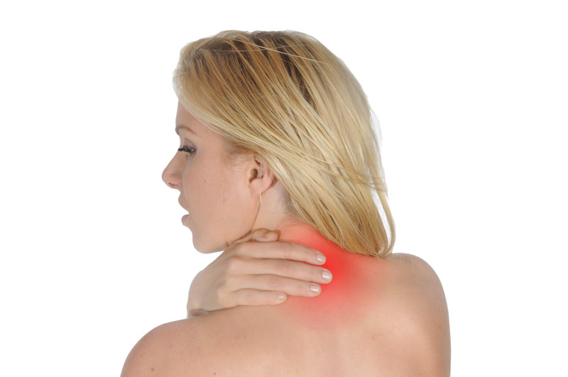
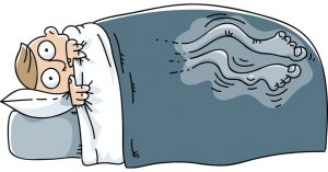
Restless legs syndrome(RLS) is a nervous system disorder resulting in an urge to move the legs. Because it usually interferes with sleep, it also is considered a sleep disorder. The condition causes an uncomfortable, “itchy,” “pins and needles,” or “creepy crawly” type feeling in the legs. These sensations are usually worse at rest, especially when lying or sitting. The symptoms are generally also worse in the evening and at night. The severity of RLS symptoms varies from mild to intolerable. Symptoms can come and go and vary widely in severity. For some people, symptoms may lead to severe sleep disturbance at night. This can significantly impair their quality of life.


Who Gets Restless Legs Syndrome?

It is estimated that ”Restless legs syndrome” (RLS) affects up to 10% of the population. It affects both sexes, but is more common in women. The condition may begin at any age, but most people affected are middle-aged or older. RLS is often unrecognized or misdiagnosed. This is especially true if the symptoms are intermittent or mild.


Causes of Restless Legs Syndrome

In most cases, doctors do not know the cause of restless legs syndrome. It is suspected that genetics plays a role. Nearly half of people with RLS also have a family member with the condition. Factors other than genetics are associated with the development of restless legs syndrome. Certain chronic diseases and medical conditions can cause RLS. These include iron deficiency, Parkinson’s disease, kidney failure, diabetes, and peripheral neuropathy. Treating these conditions often gives some relief from RLS symptoms.


Medications

Some types of medications may exacerbate symptoms. These include anti-nausea drugs, antipsychotic drugs, some antidepressants, cold and allergy medications containing sedating antihistamines. Women can experience RLS during pregnancy, especially in the last trimester. These symptoms usually go away within a month after delivery. Other factors, including alcohol use and sleep deprivation, may trigger symptoms or make them worse.


Diagnosis

There is no medical test to diagnose RLS. Doctors. However, blood tests or other diagnostic tests may be used to rule out other conditions. The diagnosis of RLS is based mainly on a patient’s symptoms and history .


Treatment

Treatment of RLS is targeted at easing the symptoms. In people with mild to moderate restless legs syndrome, lifestyle changes, such as beginning a regular exercise program , establishing regular sleep patterns, and eliminating or decreasing the use of caffeine,alcohol and tobacco, may be helpful.

Non-drug RLS treatments may include:

Leg massages

Hot baths or heating pads or ice packs applied to the legs

Good sleep habits

A vibrating pad called Relaxis


Drugs

Medications may sometimes be helpful in treatment of RLS but results vary among individuals. Drugs used to treat RLS include:

Dopaminergic drugs, which act on the neurotransmitter dopamine in the brain; Mirapex, Neupro, and Requip, levodopa.

Benzodiazepines, a class of sedative medications, may be used to help with sleep, but they can cause daytime drowsiness.

Narcotic pain relievers may be used for severe pain.

Anticonvulsant drugs  such as Tegretol, Lyrica, Neurontin,  and Horizant.


Although there is no cure for restless legs syndrome, current treatments can help control the condition, decrease symptoms, and improve sleep.


For more information see these videos     video 1.           video 2.


Restless legs syndrome treatment  by physical therapist in Tralee, Co. Kerry. Phone Eddie on 086-7700191. Click for Website

Early referral – Why it is so important – Physio in Tralee

PhysioYou have been getting treatment for back pain from your physio, chiropractor or osteopath, twice a week for the past four weeks. Each time you lie there for the first fifteen minutes with a hot pack while somebody else is being treated at the same time. The therapist pops in, has a brief chat, does a quick manipulation, reassures you of your improvement, then books you in for your next appointment. You leave wondering if you are really getting any better, but console yourself with the fact that the practitioner told you that you are.

Recently a client told me a story of where they endured a situation like the above for six months. Finally they decided to go to a doctor, who referred them for an MRI. The results showed a stress fracture to one of the lumbar vertebrae of the spine. In this case the treatments had been exacerbating the condition, and what was needed was rest and immobilisation.

Spinal Manipulations

Also manipulations, generally speaking, need to be done only once. They are used mainly to open a locked joint. A decent amount of soft tissue and myofascial work should be done by a physio beforehand to open up and relax the area. Otherwise the joint may revert to its locked position again shortly after the physio has manipulated it. Also as a general rule there should be a noticeable improvement in a clients condition from physio treatment to physio treatment. To illustrate the importance of early recognition and referral by your physio, let us look at a few more sinister conditions that present as back pain, requiring referral to a doctor or specialist.

Differential Diagnosis

Spondylitis; Ankylosing spondylitis is a condition where there is chronic inflammation of the spine and sacroiliac joints. This causes pain and stiffness in and around the spine, including the neck and back. Over time this condition can lead to a complete cementing together (fusion) of the vertebrae, a process referred to as ankylosis . Ankylosis causes loss of mobility of the spine.

Spondylolysis; A common cause of low back pain in adolescent athletes. It can be seen on X-ray and is a stress fracture in one of the bones (vertebrae) that make up the spinal column. It usually affects the fifth lumbar vertebra in the lower back, and less commonly the fourth. If the stress fracture weakens the bone too much the vertebra can start to shift out of place. This condition is called spondylolisthesis.

Spondylolysthesis; Spondylolisthesis is a condition whereby one of the vertebra of the spine slips forward or backward on the next vertebra. Spondylolisthesis can lead to deformity of the spine as well as a narrowing of the spinal canal (central spinal stenosis) and compression of the exiting nerve roots (foraminal stenosis). Spondylolisthesis is more common in the lower back but can also occur in thoracic and cervical spine.

Arthritis ; various types including spondylitis, reactive arthritis, osteoarthritis, juvenile onset spondyloarthritis, enteropathic arthritis, rheumatoid arthritis, polymyalgia rheumatica etc. can all present as back pain.

kidney stones ; can cause back pain

.Osteoporosis ; Osteoporosis means porous bones. It is a silent disease that usually goes undiagnosed until a bone fracture occurs. Bone is a living tissue that is constantly being turned over. Bones need normal sex hormones, calcium, vitamin D, adequate calories, proteins and weight bearing/strengthening exercise to keep them healthy. As we get older, more bone is lost than is replaced, but people with Osteoporosis lose more bone than people who do not have the disease. This causes bones to become more fragile and break or fracture more easily.

Various cancers ; pancreatic, liver cancers etc. can cause back pain.

ovarian cysts ; Ovarian cysts are fluid-filled sacs or pockets within or on the surface the female ovary. A large ovarian cyst can cause abdominal discomfort and a dull ache that radiates into the lower back and thighs.

Spinal stenosis ; This is a narrowing of spaces in the spine causing pressure on the spinal cord and nerves. About 75% of cases of spinal stenosis occur in the low back. In most cases, the narrowing of the spine associated with stenosis compresses a nerve root, which can cause pain down the leg.

physio in Tralee

Basically what I am saying in this article is that if your condition is not improving from physio session to physio session you may need to go back to your doctor for further investigation. Just keep it in mind. A good physio will probably have already referred you.

thoracic outlet syndromeThoracic outlet syndrome is a disorder that occurs when the blood vessels or nerves in the space between the collarbone and first rib (thoracic outlet) become compressed. This can result in any of the following symptoms ; 1. pain in your shoulders and/or neck , 2. numbness / pain / tingling down the arm and / or in a finger / fingers. Common causes include physical trauma, i.e. car crash ; repetitive injuries from job or sports related activities; anatomical defects (such as having an extra rib); poor posture( protruding neck / rounded shoulders); and pregnancy.

You may notice symptoms of thoracic outlet syndrome if your job requires you to do a repetitive movement continuously for long periods, i.e. typing on a computer; working on an assembly line; or lifting things above your head. These repetitive activities tend to tighten up neck and shoulder muscles. Athletes, such as baseball pitchers and swimmers can also develop thoracic outlet syndrome from the years of repetitive movements.

Obesity can put an undue amount of stress on your body structures and joints leading to the development of thoracic outlet syndrome. Carrying around an over sized bag or backpack or having somebody sitting on your shoulders for long periods, say at a concert can compress nerve structures etc. and lead to the condition. You may suffer symptoms during pregnancy as muscles, ligaments, tendons and joints come under strain. This is as a result of the release of the hormone relaxin around week twelve of pregnancy, and also due to the increase in bodyweight.

The two most common types of thoracic outlet syndrome are:

1. Neurogenic (neurological) : This form of thoracic outlet syndrome is characterized by compression of the brachial plexus ( nerves coming from the spinal cord (neck). This network of nerves controls muscle movements and sensations in the shoulder, arm and hand. This is the most common cause of thoracic outlet syndrome.

Signs and symptoms include:
• Numbness or tingling in your arm or fingers.
• Wasting of muscles through which the compressed nerve travels; either in the arm or fingers or both.
• Pain in the neck , shoulder or hand.
• Weakened grip in hand / loss of strength in the arm.

Treatment : Often if neurogenic thoracic outlet syndrome is caught early, deep tissue work on neck, shoulders and arm to loosen out tightened muscles; joint mobilisations / manipulation of the cervical and thoracic spine; electro acupuncture along the affected nerve pathway; postural correction exercises, can usually bring great relief within one or two treatment sessions. If the condition is chronic ( i.e. there a month or months), it may take more time for the irritated nerve to settle. You may need to lose weight if obesity is the cause of your thoracic outlet syndrome.

2.Vascular (venous or arterial): This type occurs when one or more of the veins or arteries are compressed in the area between the collarbone and first rib.
Signs and symptoms include:
• Discoloration of the hand (bluish colour).
• Arm pain and swelling, possibly due to blood clots.
• Blood clots in veins or arteries in the upper area of your body.
• Lack of colour in one or more of your fingers or your entire hand.
• Weakened or no pulse in the affected arm.
• Cold fingers, hands or arms.
• Arm fatigue after activity.
• Numbness or tingling in your fingers.
• Throbbing lump near your collar bone.
• Weakness of arm or neck.

Treatment : For vascular (venous or arterial) thoracic outlet syndrome…you need to consult with your doctor. This condition may require thrombolytic medications, anticoagulant medications or surgery.


Back Pain Treatment Options Made Simple

Three very common causes of back pain presenting in clinic are 1. muscular, 2. discogenic (from a disc), 3. locked facet joint. We will look at each and the relevant back pain treatment options.

Muscular back pain

For the low back a common muscle to cause trouble is the piriformis muscle (see diagram). It is involved in hip rotation movements so if it tightens you lose rotation putting pressure on the lower back during activities. When the piriformis muscle tightens it may also squeeze on the sciatic nerve which travels from the lumbar spine  down the back of the leg causing pain anywhere along the nerve path(low back, hamstring, calf, sole of foot). Tightened hamstrings may also contribute to low back pain(often resulting from being seated for long periods) making the spine do more work due to resulting poor posture in lifting etc.

The piriformis muscle can usually be worked out and loosened  with deep tissue massage.  The hamstrings can also be worked if these are contributing. Electro-acupuncture also helps loosen the muscles/trigger points and settle nerve irritation in affected areas.

Back pain

Locked Facet joint back pain

This is a common cause of back pain in the upper thoracic region(along back of the rib cage)…Often the person feels like somebody is sticking a finger into their back when they inhale deeply. Also with this condition a person may find that they can turn completely in one direction but not the other. In more serious cases pain may shoot down the arm due to nerve entrapment/irritation. A locked facet joint may also occur in the lower back sending nerve pain down the leg. The crack you hear when the back is manipulated is locked facet joints opening, it is not a ”slipped disc” being put back into place. A timely spinal manipulation done correctly, with a little soft tissue work done thereafter can often sort this condition. With the manipulation you get a little crack if the facet joint was locked, as it opens, giving immediate relief.

Back pain


Discogenic (Disc) related back pain

This is a more tricky one. Probably most common in the lower back (lumbar region). The patient will sometimes present with their spine all twisted off to one side in a sort of ”S” shape. This is usually because the disc bulge or protrusion is touching a nerve. The body compensates for this by trying to get the disc away from the nerve, hence the ”S” shape. Also there will be muscle spasm as the body tries to protect itself. A patient with this condition is usually in a lot of pain and even simple tasks like walking can be excruciatingly painful. Disc pain is less common in the thoracic region but again here it presents with deep unrelenting pain.

There is no such thing as a ”slipped disc”, it is just a misnomer. Discs basically bulge, degenerate(collapse) or herniate(a little fluid filled sack can protrude or leak out of them)(see diagram below). It is when this bulge or protrusion touches a nerve, the trouble starts.

Basically a muscle relaxant (valium) from your doctor along with an anti-inflammatory (NSAIDS) for a couple of days usually helps a lot.  Usually two treatment sessions along with the medication to loosen out the muscles will also reduce pain and improve recovery time. A manipulation to open any locked facet joints may help as the condition settles.

When a disc touches a nerve it initiates a series of reactions including muscle spasm and inflammation. The medication is very important in this condition and the combination of the both muscle relaxant and anti-inflammatory seem to complement each other along with treatment. Things such as ”Mckenzie technique” and ”nerve glides” can also help when introduced at the right time during treatment but require too much detail to go into here.

Once the condition settles it is important to 1. Train your body to engage its core. 2. Strengthen your core muscles with pilates type exercises, along with developing your core fitness. 3. Lose weight if you are overweight. 4. Try and improve your flexibity.

Once you get somebody moving, loosened out, give them a proper rehab routine and they are taking the medication, it is more about giving the body time to heal itself than anything else. Basically what you are trying to do with initial treatment is to relax any muscle spasm, reduce inflammation, reduce pressure on the disc with the more long-term goal of strengthening the body in the hope of bringing back in the disc bulge/ protusion enough from the nerve it is touching, so that symptoms subside. If there is little improvement in your condition after a week, an mri may be required to get more accurate information on the injury.  In some cases an epidural or surgery  may be required.

Back pain

Please note the above article only lists some common causes of back pain.

Back pain treatment in Kerry –  phone 086-7700191