Bunions

Bunions develop slowly. The big toe is made up of two joints. A bunion is a painful bony bump, also known as hallux valgus that develops on the inside of the foot at the big toe joint.  Pressure on the big toe joint causes it to bend inwards towards the second toe. Over time, the normal structure of the joint changes, resulting in a bunion. The enlarged joint is often inflamed. The deformity gradually increases making it painful to wear shoes or walk. Bunions are more common in women. Many women wear tight, narrow shoes that squeeze the toes together. This makes it more likely for a bunion to develop. In most cases, bunion pain is relieved by wearing wider shoes with adequate toe room and using other simple treatments to reduce pressure on the big toe.

Bunion Progression

An advanced bunion can greatly alter the appearance of the foot. In severe bunions, the big toe may angle all the way under or over the second toe. Pressure from the big toe may force the second toe out of alignment, causing it to come in contact with the third toe. Calluses may develop where the toes rub against each other, causing additional discomfort and difficulty walking.

Foot Problems Related to Bunions

In some cases, an enlarged big toe joint may lead to bursitis, a painful condition in which the fluid-filled sac (bursa) that cushions the bone near the joint becomes inflamed. It may also lead to chronic pain and arthritis if the smooth articular cartilage that covers the joint becomes damaged from the joint not gliding smoothly.

 Causes

Bunions may be caused by:

  • Wearing poorly fitting shoes—in particular, shoes with a narrow, pointed toe box that forces the toes into an unnatural position
  • Heredity—some people inherit feet that are more likely to develop bunions due to their shape and structure
  • Having an inflammatory condition, such as rheumatoid arthritis, or a neuromuscular condition, such as polio.

Diagnosis of bunions

Physical examination of bunions

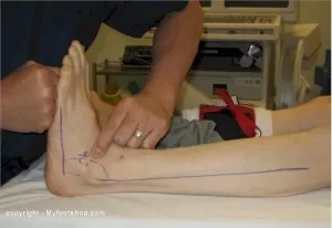
Your doctor will ask you about your medical history, general health, and symptoms. He or she will perform a careful examination of your foot. Although your doctor will probably be able to diagnose your bunion based on your symptoms and on the appearance of your toe, he or she will also order an x-ray.

X-Rays

X-rays provide images of dense structures, such as bone. An x-ray will allow your doctor to check the alignment of your toes and look for damage to the MTP joint.

Nonsurgical treatment of bunions

In most cases, bunions are treated without surgery. Although nonsurgical treatment cannot actually “reverse” a bunion, it can help reduce pain and keep the bunion from worsening.

Changes in Footwear

In the vast majority of cases, bunion pain can be managed successfully by switching to shoes that fit properly and do not compress the toes.

Padding

Protective “bunion-shield” pads can help cushion the painful area over the bunion. Pads can be purchased at a drugstore or pharmacy. Be sure to test the pads for a short time period first; the size of the pad may increase the pressure on the bump. This could worsen your pain rather than reduce it.

Orthotics and Other Devices

Orthotics (custom-made shoe inserts) may be used to take pressure off your bunion. Toe spacers can also be placed between your toes to try and straighten the big toe. In some cases, a splint worn at night that places your big toe in a straighter position may help relieve pain.

Icing

Applying ice several times a day for 20 minutes at a time can help reduce swelling. Do not apply ice directly on your skin.

Medications

Nonsteroidal anti-inflammatory medications such as ibuprofen  can help relieve pain and reduce swelling. Other medications can be prescribed to help pain and swelling in patients whose bunions are caused by arthritis.

Bunions and surgery

Your doctor may recommend surgery for a bunion, after a period of time, if you still have pain and difficulty walking despite changes in footwear and other nonsurgical treatments. Bunion surgery realigns bone, ligaments, tendons, and nerves so that the big toe can be brought back to its correct position.


Physiotherapists in Tralee stocking a wide range of orthotics to treat various foot conditions. Phone 0867700191


equinus

Equinus Foot Condition

The ankle joint connects the leg to the foot. It is formed by three separate bones, the tibia, fibula and talus. The shinbone (tibia) supports most of a person’s weight when standing. The outer bone (fibula) is the smaller bone of the lower leg. A small, irregular-shaped foot bone (talus) connects the tibia and fibula. Acting as a hinge, these bones form the ankle. The ankle joint allows movement such as walking, running and jumping, and also contributes to lower limb stability.

The ankle is reinforced by ligaments which connect bone to bone. Ligaments have a mildly elastic structure that allows them to stretch, within their limits, and then return to their normal positions. Ligaments protect the ankle from abnormal movements—especially twisting, turning and rolling of the foot.

Description

A person with equinus has a limited range of ankle motion and lacks the flexibility needed to bring the top of the foot upward, toward the shin. It may be either congenital or acquired, and occurs equally in both men and women.  Equinus can be due to several different reasons including the following:

• bony block between the talus and distal tibia (osseous equinus);
• contracture or tightness of the soleus muscle (inner calf muscle);
• contracture or tightness of the soleus and gastrocnemius muscles (gastroc-soleal equinus);
• isolated tightness of the gastrocnemius muscles (outer calf muscle); and
• compensatory loss of ankle joint range of motion for some other condition such as pes cavus (pseudoequinus).


People with equinus often develop ways to compensate for their limited ankle motion. Depending on how a patient compensates for the inability to bend properly at the ankle, a variety of other foot conditions can develop, such as:

  • Plantar Fasciitis
  • Calf cramping
  • Achilles Tendinitis
  • Metatarsalgia (pain and/or callusing on the ball of the foot)
  • Flatfoot
  • Arthritis of the midfoot (middle area of the foot)
  • Pressure sores on the ball of the foot or the arch
  • Bunions and hammertoes
  • Ankle pain
  • Shin splints
  • Sesamoiditis
  • Hallux valgus
  • hallux rigidus
  • Hammer toes


Nonsurgical Treatment of Equinus

Some nonsurgical treatment strategies are aimed at relieving the symptoms and conditions associated with equinus. Treatment for the equinus itself may include one or more of the following options.

  • Heel lifts—Placing heel lifts inside the shoes or wearing shoes with a moderate heel may reduce symptoms by taking stress off the Achilles tendon and compensating for the restricted movement of the ankle joint. The joint is meant to have about fourteen degrees of movement ideally, so say it has only four degrees of movement, adding a ten degree heel lift helps compensate for the missing degrees of movement .
  • Arch supports or orthotic devices—Custom orthotic devices that fit into the shoe are often prescribed to ensure that weight is distributed properly, and to help control muscle/tendon imbalance. Again these devices will most likely include a heel lift.
  • Physical therapy—To help remedy muscle tightness, deep tissue massage of calf muscles along with a stretching program for the calf muscles are recommended.


For a quick simple video describing the condition click   here


See our wide range of orthotics  here


What is Hallux Rigidus?

Hallux Rigidus

Big toe joint arthritis (otherwise known as Hallux Rigidus) is a form of degenerative arthritis. In this condition, surfaces of the joints in the big toe begin to wear away and extra bone can also develop in the form of  bone spurs or osteophytes. This limits the movement of the joint. The big toe  needs to bend significantly when stepping off. Consequently, arthritis in this joint can greatly affect walking, running etc.

Hallux Rigidus is a condition that tends to get worse over time. In it’s earlier stages, it may be referred to as Hallux Limitus, which is where there is limited movement of the big toe joint. With progression, Hallux Rigidus develops which can result in stiffness developing in the big toe joint and there may also be swelling.

To compensate for pain in the big toe joint, people with Hallux Rigidus tend to adjust their walking pattern which can also result in knee, hip or lower back pain. There is often difficulty finding shoes that fit properly shoes due to pain and/or inflammation in the joint. Early diagnosis can be made by physical examination and x-ray. Early treatment gives the best chance of avoiding surgery.

What causes Hallux Rigidus?

The big toe joint is designed to bear a considerable amount of stress during walking. However faulty biomechanics or structural abnormalities such as flat feet can stop the big toe from bending normally, and eventually lead to the development of osteoarthritis in the big toe joint. Other factors that can increase the risk of developing this condition include genetics (eg, having a certain foot type), injury to the big toe , other inflammatory diseases, and working in a job that places excessive stress on the big toe joint .

Treatment

In cases of Hallux Rigidus, certain types of footwear and orthotics may be recommended to reduce pressure and motion on the big toe.  Custom orthotics that conform very closely to the arch of the foot are most effective in improving big toe joint function. These orthotics may incorporate  something like a 3mm kinetic wedge in combination with a built in forefoot posting to take the pressure off the big toe joint. Ice and anti-inflammatory medication can also be used to reduce inflammation and pain. If it does not respond to conservative treatment, surgery may be recommended as a last resort in more severe cases.

physiotherapist
Fig. 1. pronation-supination

Physiotherapist explains foot pronation & supination

A physiotherapist explains Pronation and supination as  movements that occur at the subtalar joint of the foot. The normal biomechanics of the foot are designed to absorb and direct the forces occurring throughout the gait cycle. As the foot is loaded, eversion of the subtalar joint, dorsiflextion of the ankle, and abduction of the forefoot occur. This is the pronation part of the gait cycle. Pronation should not continue past the latter stages of midstance during the gait cycle. At this stage the foot should then supinate in preparation for toe-off. Approximately four degrees of pronation and supination are necessary to enable the foot to propel forward properly. Any increase on this four degrees brings a foot into over-supination or over-pronation.


Pronation – physiotherapist explaination

Pronation of the foot is where the heel and the little toe move away from the center of the body. The foot also dorsiflexes up slightly, the ankle rolling inwards. Pronation is part of the natural movement of the human body. Certain injuries can occur with excessive pronation.  Runners with flat feet often tend to overpronate. Over-pronation can contribute to many injuries. These include shin splints, anterior compartment syndrome, patello-femoral pain syndrome, plantar fasciitis, tarsal tunnel syndrome, bunions (hallux valgus), achilles tendonopathies etc. The running shoes of over-pronators often show extra wear on the inner heel and ball of the foot.


Supination – physiotheraist explaination

With supination the heel and also the big toe rotate towards the centre of the body. The foot flexes down and the ankle rolls out. It is the opposite of pronation. A natural amount of supination occurs during the push-off phase of the running gait. This occurs as the heel lifts off the ground and the forefoot and toes are used to propel the body forward. However, excessive supination  places a large strain on the muscles and tendons that stabilize the ankle. This can make the oversupinator more prone to ankle sprain or ankle ligament rupture.

With over-supination the forces of impact on the foot are concentrated on a smaller area of the foot (the outside part), and are not distributed as efficiently. In the push-off phase, most of the work is done by the smaller toes on the outside of the foot, rather than the big toe. This places extra stress on the foot. It can lead to conditions such as iliotibial band syndrome, Achilles tendinitis, or plantar fasciitis. Over-supination causes the outer edge of running shoes to wear sooner. In extreme cases, there will also be holes in the uppers where the runner’s foot has broken through. Runners with high arches and tight Achilles tendons/calves tend to be over-supinators.


Orthotics

Properly prescribed foot orthotics can be beneficial in the treatment of over-pronation or over-supination. Physiotherapists usually supply both off the shelf and/or custom made orthotics. We  prescribe these only when we really feel they are necessary. Often the much cheaper off the shelf version will do the trick. It is important to get a quality product, prescribed by an experienced practitioner. Sometimes you just have an injury that needs  treatment and there is no need for an orthotic. Also high arches or a flat feet do not mean in itself you need orthotics, especially if you are not in pain.


Metatarsalgia

Metatarsalgia
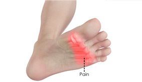
Metatarsalgia is the name given to pain in the front part of your foot under the heads of your metatarsal bones ( ball of foot, just before toes). It is usually worse when standing or walking etc.  and occurs most frequently in the second, third/or fourth metatarsal joints or isolated in the first metatarsal joint. Metatarsalgia usually comes on gradually over some weeks rather than suddenly. The affected area of your foot may also feel tender on palpation by your physiotherapist.

Common causes identified by physiotherapists include:

  • Pes cavus or high arched foot.
  • Excessive pronation of the foot(foot rools inwards ie. with flat foot).
  • Clawing or hammer toes.
  • Tight extensor tendons of the toes.
  • Prominent metatarsal heads.
  • Morton’s foot – here there is a shortened first metatarsal, which results in an abnormal gait putting increased pressure on the second metarsal.
  • Over doing it in athletes such as runners and tennis players etc. can lead to inflammation in the joints due to the pounding they receive.
  • Being overweight puts extra stress on many areas of the foot including metatarsals.
  • Wearing high heels – forward force on feet increases the pressure on metatarsal area.
  • Having tightened calf muscles or poor ankle flexibility – this affects the normal gait pattern.
  • Poorly fitting or tight footwear can squeeze the metatarsal joints causing friction , inflammation and poor gait.

Simple measures can help to relieve the symptoms of metatarsalgia. These include:

  • Resting with your feet elevated where possible.
  • Losing weight if you are overweight.
  • Wearing supportive shoes that are well fitted, low-heeled and have a wide toe area.
  • Metatarsal pads and orthotic inserts for your shoes may help to relieve pain in your forefoot by reducing the pressure placed on the heads of your metatarsal bones.
  • Physiotherapy may also be helpful. This may include stretching out the spaces between the metatarsal and mobilising the joints of the foot or performing deep tissue massage to loosen your calf muscles or any other lower limb muscles that may be contributing to poor gait. Sometimes if a metatarsal head has dropped it can be manipulated back into place. Pain relief as a result of this mobilisation, when effective can be immense and long lasting.
  • Simple painkillers such as paracetamol and non-steroidal anti-inflammatories(NSAIDs) such as ibuprofen may help to relieve pain.