thoracic spine mobility
Thoracic Spine

A mobile thoracic spine allows you turn your back in many directions, enabling you to do everyday tasks with ease. Today’s sedentary lifestyle often contributes to reduced spinal mobility. Basically ”motion is lotion” and if you are inactive and also prone to poor posture, your thoracic spine can seize up. If it goes on long enough, say into old age portions of the spine may fuse and not move at all. A lack of thoracic spine mobility means that the lumbar spine, pelvis, shoulders and surrounding muscles  have to compensate. Long term, these over-compensations can  lead to overuse conditions and injuries, the lower back being particularly suceptable. This is because the lumbar spine is meant to keep us stable and is not very mobile, so when these joints are forced to overcompensate for the lack of movement in the thoracic area, it can place alot of pressure on the discs of the lower back. Possible consequences include inflammation, degeneration,  herniation of the discs, generalized low back pain, compression fractures, muscles spasms, and spinal nerve injuries. Similar pressures and injuries can occur in the neck and shoulders. For example, if your thoracic spine isn’t mobile, anytime you have to do a movement overhead, your shoulders make up for that lack of mobility. If you have shoulder impingement or chronic shoulder and neck problems lack of mobility in the thoracic spine will make every thing worse.


Improving Thoracic Spine Mobility

Yoga, pre- and post-workout stretching, and mobility exercises are the best way of maintaining and improving thoracic spine mobility. These need to be done regularly and consistently, especially as you get older. Your physiotherapist will be able to guide you on the correct exercises and help correct your form and technique until you get used to them and can do them yourself. Here are a few exercises to get you started.

For more information contact us @ Physiotherapists in Tralee.


Back Mice
Fig. 1 Back Mice

The term “back mice” is a rather cute description for a painful yet often overlooked condition, even by back pain specialists. The term  “back mice” was first used to label the condition by Peter Curtis in 1993. Back mice present as small, firm, fleshy yet moveable nodules upon palpation over the sacral region. Firm pressure directly on the nodules usually produces pain and tenderness, which sometimes radiates into the sacrum and hip. Also, the back mouse seems to suddenly appear following trauma to the back as in a motor vehicle accident or perhaps following a lifting injury. The size of the nodules does not change and they remain the same regardless of the administered soft tissue treatment.

Perhaps a more descriptive term for the back mouse is  “lumbar fascial fat herniation” . This occurs when the lumbar subfascial fat layer herniates through the overlying thoraco-dorsal fascia and gets trapped and inflamed. The mechanism appears to be due to an anatomical defect or weakened area in the fascia, which, when there is increased internal pressure, allows the fat lobules to push through the fascia. Once herniated, the fat becomes trapped and as an expanded, inflamed, herniation in an otherwise unyielding fibrous capsule. This creates a focus of pain. Pressure on the fat mouse does not push it back through the fascia but only inflames the torn fascia more. These herniations occur at predictable sites along the iliac crest and sacrum very close to the natural dimple area. They also are approximately three times more prevalent in women, particularly in moderately obese women.

A client usually presents with an episode of  low back pain. There may be a history of pain with lifting or prolonged sitting and the pain is usually greater on one side more than the other. The pain may radiate into the buttocks and sacrum and perhaps to the lateral thigh and into the lower extremity. Medications usually do nothing. Many will have gone through the pain pill merry-go-round, taking a cocktail of pain and anti-inflammatory medications even though the pain never truly goes away. The client has often tried everything, been everywhere and you are their last hope. They may have had radiographs, MRI studies or nerve conduction studies, all with negative or minimal findings. They may even have a minor disc bulge without nerve compression, yet the pain exactly mimics a discogenic disorder. When asked to point to the area of the greatest pain, they will invariably point just above and lateral to the natural “dimple” where the back and buttocks come together, near the multifidis triangle. When the area is palpated, the most marked finding is one or several firm, mobile 1.3 cm nodules. When pressed, these nodules reproduce the client’s complaints of back pain as well as the “sciatic” pain.

Subcutaneous lipomas in the back region differ from back mice in that they present as moveable non-tender “speed bumps” that cause pain only when they compress the underlying soft tissue. Subcutaneous lipomas can be found anywhere in the body. They grow slowly over time and are only cosmetically important.

back mice
Fig 2. Overlying thoraco-dorsal fascia.


back mice
Fig 3. Iliac crest and sacrum.


There always seems to be a focus by back pain specialists upon the disc and nerves issues even though the fat mice are readily palpable. Many sufferer’s have had epidural injections without success. I have had clients who have had surgery for disc herniations yet who still point to the back mouse post surgery as the focal point of pain. It could perhaps be considered diagnostic for the presence of a lumbar fascial fat herniation if a local infiltration of anesthetic takes away the pain.

Treatment of Back Mice

A medical doctor can inject the back mouse with a local anesthetic. This usually only helps temporarily. Dry needling techniques by acupuncturists may help reduce the tension in the fibrous capsule. Good results may sometimes be obtained with local electrical stimulation techniques such as electro-acupuncture. Do not apply deep pressure to back mice during manual physiotherapy treatment. Doing so may only serve to aggravate the herniation. Release of muscular tissue tension around the back mouse may provide some relief by easing pressure on the area.

Perhaps the only permanent cure for the back mouse is its excision and removal. This could be performed by a hernia repair specialist. Once the fat herniation is excised and the fascial tear repaired, the client usually enjoys a more enduring and sometimes dramatic relief. One of the biggest problems is that so many medical doctors fail to recognize this condition; they tend to discount its existence, thereby limiting the treatment options. Icing the area may also provide temporary relief.


For a short video on back mice click here.


Physiotherapist in Tralee. Phone 0867700191 to discuss your injury, make an appointment or get a second opinion.


Achilles tendon ruptures will debilitate your lower leg completely, the moment they happen. The Achilles tendon is an important part of the leg. It is located just behind and above the heel, attaching the heel bone to the calf muscles. Its function is to help in bending the foot downwards at the ankle (this movement is called plantar flexion). If the Achilles tendon is torn/ruptured, the tear may be either partial or complete. In a partial tear, the tendon is torn but is still partly joined to the calf muscle. With complete tears, the tendon is completely torn and the connection between the calf muscles and the ankle bone is lost. If your Achilles tendon is ruptured you will be unable to stand on your tiptoes and you will have a flat-footed walk with a severe limp, along with considerable pain initially.


An Achilles tendon can tear when there is too high a load or stress placed on it. This can happen with activities involving a forceful push off with the foot – ie. running/sprinting in football, basketball, tennis etc. I have also seen it occur when somebody sprints off suddenly on a sandy beach, the sand giving way under the foot putting extra stress on the calf and the achilles tendon. The push off movement uses a strong contraction of the calf muscles maximally stressing both the calf muscles and the Achilles tendon . Injury to the achilles can also occur due to falls, if the foot is suddenly forced into an upward-pointing position, stretching the tendon. Another possible injury is a deep cut/laceration to the tendon.


Achilles Tendon Rupture
   Fig. 1.  Achilles Tendon Rupture


Sometimes the Achilles tendon is weak, making it more prone to rupture. Factors that weaken the Achilles tendon are:

  • Corticosteroid medications (such as prednisolone) – mainly when used as long-term treatment.
  • A corticosteroid injection near the Achilles tendon.
  • Certain rare medical conditions, such as Cushing’s syndrome, where the body makes too much of its own corticosteroid hormones.
  • Tendinopathies of the Achilles tendon.
  • Other medical conditions which can make the tendon more prone to rupture; for example, rheumatoid arthritis, diabetes, gout and systemic lupus erythematosus.
  • Certain antibiotic medicines may slightly increase the risk of having an Achilles tendon rupture. These are the quinolone antibiotics such as ciprofloxacin and ofloacin. The risk of having an Achilles tendon rupture with these antibiotics is quite low and mainly applies to people who are also taking corticosteroid medication.


You might hear a snap or feel a sudden sharp pain when the tendon is torn (ruptured)during a sporting activity or injury. The sharp pain usually settles quickly, although there may be some aching at the back of the lower leg. After the injury, the usual symptoms are:

  • A flat-footed type of walk. You can walk and bear weight but cannot push off the ground properly on the side where the tendon is ruptured.
  • Inability to stand on tiptoe.
  • If the tendon is completely torn, you may feel a gap just above the back of the heel. However, if there is bruising and swelling may disguise the gap.

An Achilles tendon rupture is usually diagnosed based on symptoms, history of the injury and a physio/doctor’s examination. An orthopedic test called Thompson’s test (also known as the calf squeeze test) may help diagnosis. In this test, you will be asked to lie face down on the examination bench and to bend your knee. The doctor will gently squeeze the calf muscles at the back of your leg and observe how the ankle moves. If the Achilles tendon is OK, the calf squeeze will make the foot point away from the leg (a movement called plantar flexion). This is quite an accurate test for Achilles tendon ruptures. An ultrasound or MRI may be used to confirm diagnosis and give a fuller picture of the injury.


Treatment and recovery

Treatment for a ruptured achilles tendon usually involves surgery to repair the tendon. The surgeon sews/sticks together the torn ends of the Achilles tendon, and may also use another tendon or a tendon graft to help with the repair. A plaster cast or brace is needed after the operation for about eight weeks, to keep the foot immobilized, allowing the tendon can heal. The plaster cast or the brace is positioned so that the foot is pointing slightly downwards, which takes the strain off the tendon.

Traditionally, crutches were used to keep weight off the leg during the first few weeks of treatment. Current thinking tends towards using the leg normally early on (early mobilization). This involves fitting a plaster cast or a brace which you can walk on. It is more convenient because you do not need to use crutches. Physiotherapy will also be needed, especially when the cast is removed. I have personally found instrument assisted soft tissue work(after about 8 weeks) to be very beneficial along with stretching/strengthening work and deep tissue work to the calf during recovery. Full recovery is greatly slowed when there is no hands-on work done during recovery, after the cast is removed once an ok is given by the surgeon to commence.

Depending on a person’s profession, some people may need several weeks off work after an Achilles tendon tear (rupture); the time taken to return to sport is usually between 4 and 12 months. Generally, the outlook is good. However, the tendon does take time to heal, usually about six to eight weeks. More time will be needed after this to allow the muscles and tendon to regain normal strength.


For more on rehabilitation on achilles tendon ruptures post surgery click here.


Physiotherapist in Tralee. Ring 08677001 to discuss your condition, book an appointment or get a second opinion.

Meniscus Injuries  – Knee Pain

Meniscus injuries are a relatively common knee injury. Cartilage within the knee joint provides cushioning between the bones at this joint. This protects them from the stresses of walking, jumping, running etc. There is articular cartilage which is the smooth, white tissue that covers the ends of bones(Femur, tibia) where they come together to form the knee joint. Healthy cartilage in our joints makes it easier to move. It allows the bones to glide over each other with very little friction. Articular meniscus injuries
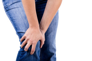
cartilage can be damaged by injury or normal wear and tear.

Within the knee you also have fibrocartilage in the form of the medial and lateral meniscii. These are two thick wedge-shaped pads of cartilage attached to top of the tibia (tibial plateau) and under the femur bone. They  allow the femur to glide when the knee joint moves. Each meniscus is curved in a C-shape, with the front part of the cartilage called the anterior horn and the back part called the posterior horn. Meniscal tears are usually described by where they are located anatomically in the C shape and by their appearance (for example, “bucket handle” tear, longitudinal, parrot beak, and transverse).

Blood supply

Because the blood supply is different to each part of the meniscus. Knowing where the tear is located may help decide how easily an injury might heal (with or without surgery). The better the blood supply, the better the potential for recovery. The outside rim of cartilage has better blood supply than the central part of the “C.” Blood supply to knee cartilage also decreases with age, with up to 20% of normal blood supply  lost by age 40.

What causes a meniscus to tear?

A forceful twist or sudden stop can cause the end of the femur to grind into the top of the tibia, pinching and potentially tearing the cartilage of the meniscus. These meniscus injuries can also occur with deep squatting or kneeling, especially when lifting a heavy weight. Meniscus tear injuries often occur during athletic activities, especially in contact sports like football and hockey. Motions that require pivoting and sudden stops, in sports like tennis, basketball, and golf, can also cause meniscus damage.


Meniscus injuries  – Increasing risk with age

The risk of developing a torn meniscus increases with age. This is  because cartilage begins to gradually wear out, losing its blood supply and its resilience. Being overweight also puts more stress on the meniscii. This means that routine daily activities like walking and climbing stairs increase the potential for wear, degeneration, and tearing. It is estimated that six out of 10 patients older than 65 years have a degenerative meniscus tear. Many of these tears may never cause problems.

Some of the fibers of the cartilage are interconnected with those of the ligaments that surround the knee. Thus, meniscus injuries may also be associated with tears of the collateral and cruciate ligaments, depending upon the mechanism of injury.

Symptoms of meniscus injuries can include some or all of the following:

  • Pain with running or walking longer distances
  • Intermittent swelling of the knee joint: Many times, the knee with a torn meniscus feels “tight.”
  • Popping, especially when climbing up or down stairs
  • Giving way or buckling (the sensation that the knee is unstable and the feeling that the knee will give way): Less commonly, the knee actually will give way and cause the patient to fall.
  • Locking (a mechanical block where the knee cannot be fully extended or straightened): This occurs when a piece of torn meniscus folds on itself and blocks full range of motion of the knee joint. The knee gets “stuck,” usually flexed between 15 and 30 degrees and cannot bend or straighten from that position.  

Diagnosis

The diagnosis of a knee injury begins with a history of the injury etc. and physical examination. There have been many tests described to assess the internal structures of the knee. The McMurray test is one long used orthopedic test . The health-care professional flexes the knee and rotates the tibia while feeling along the joint. The test is positive for a potential tear if a click is felt or noticeable pain is felt while circumducting the knee in full flexion.

Physiotherapy in Tralee – referral for MRI

(MRI) is the test of choice to confirm the diagnosis of a torn meniscus. It also allows a radiographer to visualize the inner structures of the knee. These structure include; the cartilage and ligaments, the surface of the bones, and the muscles and tendons that surround the knee joint. Plain X-rays cannot be used to identify meniscal tears but may be helpful in looking for bony changes, including fractures, arthritis, and loose bony fragments within the joint. In older patients, X-rays may be taken of both knees while the patient is standing. This allows the joint spaces to be compared to assess the degree of cartilage wear. Cartilage takes up space within the joint and if the joint space is narrowed, it may be an indicator that there is less cartilage present, likely from degenerative disease.

Treatment of Meniscal Tears

Sometimes conservative measures such as physical therapy, NSAIDs and rest can be enough to settle the condition. When conservative measures are ineffective the next step may be surgery to repair or remove the damaged cartilage.


Here is a little video explaining more.


For physiotherapy in Tralee phone 086-7700191, click here for website homepage
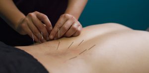
Dry-NeedlingDry Needling

Trigger-point dry needling is a procedure where a fine  acupuncture needle is inserted into the skin and muscle. It is aimed at myofascial trigger points, which are points of exquisite pain in skeletal muscle, that are associated with a hypersensitive palpable nodule or a taut band.

Active trigger points can spontaneously trigger local or referred pain. They cause muscle weakness, restricted range of movement and autonomic phenomena. Latent trigger points do not cause pain unless they are stimulated. They may alter muscle activation patterns and contribute to restricted range of movement . Therefore both active and latent trigger points cause allodynia(nerve pain) at the trigger point site and hyperalgesia away from the trigger point following applied pressure.

The formation of trigger points is caused by the creation of a taut band or knot within the muscle. This band is caused by excessive acetylcholine release from the motor endplate combined with inhibition of acetylcholine esterase and upregulation of nicotinic acetylcholine receptors.  Motor end plates, also called neuromuscular junctions, are specialised chemical synapses formed at the sites where the terminal branches of the axon of a motor neuron contact a target muscle cell. Motor neurons are nerve cells that send electrical output signals to the muscles.

Initially the taut bands are produced as a normal protective, physiological measure in the presence of actual or potential muscle damage. They are thought to occur in response to unaccustomed eccentric or concentric loading, sustained postures and repetitive low load stress. However when sustained they contribute to sustained pain. The pain caused by trigger points is due to hypoxia and decreased blood flow within the trigger point. This leads to a decreased pH which activates the muscle nociceptors to restore homeostasis. This causes peripheral sensitization. Trigger points are also involved in central sensitization. The mechanism remains unclear but trigger points maintain nocioceptive input into the dorsal horn and therefore contribute to central sensitization.


Stimulation of a local twitch response (LTR)

Dry-needling of these myofascial trigger points via mechanical stimulation causes an analgesic effect. This mechanical stimulation causes a local twitch response (LTR). A LTR is an involuntary spinal cord reflex contraction of the muscle fibers in a taut band. Triggering an LTR has been shown to reduce the concentration of nociceptive substances in the chemical environment near myofascial trigger points.


Muscle regeneration

The needle may also cause a small focal lesion which triggers satellite cell migration to the area which then repair or replace damaged myofibers. This occurs 7-10 days after dry needling. It is unclear whether continued dry needling within this period may disrupt this process.

Dry needling may also cause a localized stretch to the cytoskeletal structures. This stretch may allow sarcomeres to resume their resting length. The mechanical pressure causes collagen fibers to intrinsically electrically polarize which also triggers tissue remodeling.


The effectiveness of this treatment depends greatly on the skill of the therapist to accurately palpate mysofascial trigger points.


video on twitch response of dry needling


Physiotherapists in Tralee specializing in hand-on deep tissue work, dry needling and osteopathic manipulations. Phone 0867700191 to discuss your condition or make an appointment.

equinus

Equinus Foot Condition

The ankle joint connects the leg to the foot. It is formed by three separate bones, the tibia, fibula and talus. The shinbone (tibia) supports most of a person’s weight when standing. The outer bone (fibula) is the smaller bone of the lower leg. A small, irregular-shaped foot bone (talus) connects the tibia and fibula. Acting as a hinge, these bones form the ankle. The ankle joint allows movement such as walking, running and jumping, and also contributes to lower limb stability.

The ankle is reinforced by ligaments which connect bone to bone. Ligaments have a mildly elastic structure that allows them to stretch, within their limits, and then return to their normal positions. Ligaments protect the ankle from abnormal movements—especially twisting, turning and rolling of the foot.

Description

A person with equinus has a limited range of ankle motion and lacks the flexibility needed to bring the top of the foot upward, toward the shin. It may be either congenital or acquired, and occurs equally in both men and women.  Equinus can be due to several different reasons including the following:

• bony block between the talus and distal tibia (osseous equinus);
• contracture or tightness of the soleus muscle (inner calf muscle);
• contracture or tightness of the soleus and gastrocnemius muscles (gastroc-soleal equinus);
• isolated tightness of the gastrocnemius muscles (outer calf muscle); and
• compensatory loss of ankle joint range of motion for some other condition such as pes cavus (pseudoequinus).


People with equinus often develop ways to compensate for their limited ankle motion. Depending on how a patient compensates for the inability to bend properly at the ankle, a variety of other foot conditions can develop, such as:

  • Plantar Fasciitis
  • Calf cramping
  • Achilles Tendinitis
  • Metatarsalgia (pain and/or callusing on the ball of the foot)
  • Flatfoot
  • Arthritis of the midfoot (middle area of the foot)
  • Pressure sores on the ball of the foot or the arch
  • Bunions and hammertoes
  • Ankle pain
  • Shin splints
  • Sesamoiditis
  • Hallux valgus
  • hallux rigidus
  • Hammer toes


Nonsurgical Treatment of Equinus

Some nonsurgical treatment strategies are aimed at relieving the symptoms and conditions associated with equinus. Treatment for the equinus itself may include one or more of the following options.

  • Heel lifts—Placing heel lifts inside the shoes or wearing shoes with a moderate heel may reduce symptoms by taking stress off the Achilles tendon and compensating for the restricted movement of the ankle joint. The joint is meant to have about fourteen degrees of movement ideally, so say it has only four degrees of movement, adding a ten degree heel lift helps compensate for the missing degrees of movement .
  • Arch supports or orthotic devices—Custom orthotic devices that fit into the shoe are often prescribed to ensure that weight is distributed properly, and to help control muscle/tendon imbalance. Again these devices will most likely include a heel lift.
  • Physical therapy—To help remedy muscle tightness, deep tissue massage of calf muscles along with a stretching program for the calf muscles are recommended.


For a quick simple video describing the condition click   here


See our wide range of orthotics  here


Posterior ankle impingement physiotherapy
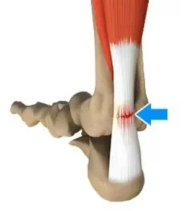
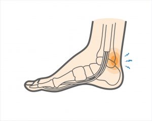
Posterior ankle impingement is a condition characterised by tissue damage at the back of
the ankle joint due to compression of these structures. This occurs when the foot and ankle
are pointed maximally away from the body (plantarflexion – figure 1. ). It may occur when
compressive forces are too repetitive and/or too forceful. This can occurs in the presence of
ankle swelling or bony anomalies, such as additional bone, a condition known as an “os
trigonum”. Posterior ankle impingement is most commonly found in gymnasts, ballet
dancers, and footballers, because they regularly maximally plantarflex their ankles during
their activities. The condition can also occur due to inadequate rehabilitation of an acute
ankle injury (ie. ankle sprain).

Mechanism of Injury

Posterior ankle impingement may develop due to an acute traumatic plantar hyperflexion
event, such as an ankle sprain. It may also occur as a result of repetitive low-grade trauma
associated with plantar hyperflexion, say like in case of a female ballet dancer. It is
important to differentiate between these two, because the latter, that is posterior
impingement from overuse, has a better prognosis.
The anatomy of the posterior ankle is a key factor in the occurrence of posterior
impingement syndrome . The more common causes of the condition are osseous in nature,
such as the os trigonum, an elongated posterolateral tubercle of the talus (known as
Stieda’s process), a downward sloping posterior lip of the tibia, an osteophyte from the
posterior distal tibia , or a prominent posterior process of the calcaneus. However, posterior
impingement can also be soft tissue related, as with a thickened posterior joint capsule ,
post-traumatic scar tissue, post-traumatic calcifications of the posterior joint capsule, or
loose bodies in the posterior part of the ankle joint. Symptoms for all of these conditions
relate to physical impingement of osseous or soft tissue structures, resulting in painful
limitation of the full range of ankle movement.
The most common cause ''os trigonum'' is an extra (accessory) bone that sometimes
develops behind the ankle bone (talus). The mineralized os trigonum appears between the
ages of 7 and 13 years and usually fuses with the talus within 1 year, forming the trigonal
(Stieda) process. It may remain as a separate ossicle in 7-14% of patients, and is often
bilateral(in both ankles). An os trigonum can be a focus of osseous abutment against other
structures. Pain can also be caused by disruption of the cartilaginous synchondrosis
between the os trigonum and the lateral talar tubercle as a result of repetitive microtrauma
and chronic inflammation.
In the case of soft tissue impingement it usually results from scarring and fibrosis associated
with synovial, capsular, or ligamentous injury ie. bad ankle sprain. It is thought that this
type of manifestation usually usually occurs when a significant soft-tissue component
forms. The soft-tissue component can consist of synovial thickening throughout the
posterior capsule or be more focal, involving the posterior intermalleolar or talofibular ligament. The flexor hallucis longus tendon runs in the groove between the lateral and
medial processes of the talus and can also be injured in posterior impingement, resulting in
tenosynovitis.


Signs and symptoms

Patients who have posterior impingement complain of chronic deep posterior ankle pain
worsened by forced plantar flexion or push-off forces as occur during activities such as
ballet dancing, jumping, or running downhill. In some patients, forced dorsiflexion(opposite
to plantarflexion) is also painful. Physical examination reveals pain on palpation over the posterolateral talar process, which is located along the posterolateral aspect of the ankle between the Achilles and peroneal
tendons . Passive forced plantar flexion results in pain and often a grinding
sensation as the posterolateral talar process is entrapped between the posterior tibia and
calcaneus.


Diagnosis of posterior ankle impingement

A thorough examination by an experienced practitioner may be all that is necessary to
diagnose posterior ankle impingement. Further investigations such as an X-ray, MRI, CT scan
or Ultrasound may help confirm diagnosis.


Physiotherapist in Tralee, Co. Kerry………..Phone 0867700191 to make an appointment or discuss your condition.

Suboccipital-Muscles-of-the-Neck

Suboccipital Muscles & Trigger Point Pain

Suboccipital muscles (see image) are a group of four muscles located on each side of the upper cervical spines, just below the base of the skull. The muscles connect the base of the skull with the top two vertebrae (C1 and C2) of the neck.

Poor posture in general especially with the increased use of portable electronic devices, which include mobile phones, laptops, and tablets has increased the prevalence of neck pain in both children and adults. Increased screen time on these devices is not only correlated with depression, sleep interruption, and poor food choices, but also rising rates of neck pain, especially in adolescents and young adults. This form of neck pain, including dysfunction of C1 and altered mechanics of the cervical spine due to poor posture, can also lead to headaches. Reading in bed is also a big offender. Any position where your head and neck are positioned forward and in a stationary position for long periods on time increases the likelihood of postural related spinal issues(ie. kyphosis, discogenic disorders etc.), neck pain and headaches.

These suboccipital muscles play an important role in controlling movements of your head and neck, providing sensory input and are also linked closely to vestibular and balance functions. However, when the suboccipital muscles become tightened, the following symptoms may occur. These could include:

  • Stiff neck
  • Neck pain
  • Headaches with a band of pain on the side of the head that extends from the back of the head to the eye as a result of active trigger points. This type of pain feels deep in the head, and often it is difficult to describe.

Messages sent to the brain may be altered, which is also why sometimes headache sufferers may also experience sensory symptoms, including dizziness and visual disturbances.


Physiotherapy Treatment

Treatment includes deep tissue work, trigger point release, manipulation, mobilisation, postural education and a rehabilitation program.


suboccipttal trigger points
Referred pain from suboccipital trigger points


Physiotherapists in Tralee Phone 0867700191


myofascial-pain-syndrome
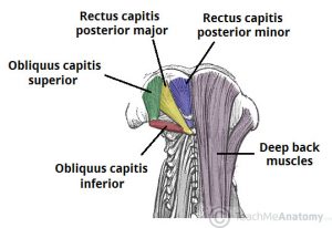
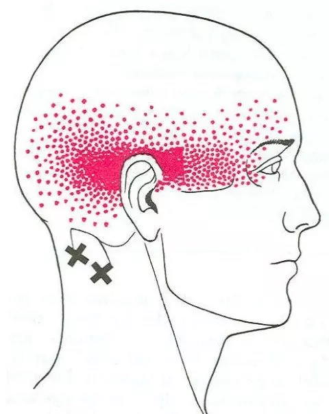
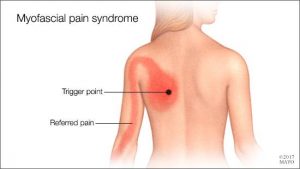
Myofascial Pain Syndrome

Myofascial pain syndrome is where pressure on sensitive points within your muscles (trigger points) causes pain and sometimes refers pain to seemingly unrelated parts of your body. It can occur after a muscle has been contracted repetitively ie. repetitive motions used in jobs or hobbies or by stress-related muscle tension. While nearly everyone has experienced muscle tension pain at some point, the discomfort associated with myofascial pain syndrome persists or worsens. There is no laboratory, radiographic or other diagnostic tests to prove the diagnosis of myofascial pain syndrome so it is considered a “subjective” diagnosis. Myofascial trigger points do not always cause pain. Sometimes they can lie dormant or inactive within a muscle for months or even years. Trigger points can often be identified by a skilled therapist working through your muscles with deep tissue massage. They feel like little knots deep within the muscles which when pressed are extremely tender.

Fascia is the body’s connective tissue. It is a head to toe, all-encompassing and interwoven system of fibrous connective tissue found throughout the body. Your  fascia provides a framework that helps support and protect individual muscle groups, organs, and the entire body as a unit. It is the same as that cling film, elastic type structure that surrounds a joint of meat, helping hold it together when the outer skin is removed.

This fascia in itself can also contribute to ”myofascial pain” syndrome. Injury, illness, stress, aging and repetitive use, can cause the fascia to shorten, thicken and become more unyielding . All the nerves and blood vessels run through the fascia. Therefore, if this connective tissue is tight, the associated tissues will have poor nutrient exchange. This exacerbates any painful situation because toxic metabolic waste products build up which often further aggravate pain receptors. This can create a vicious cycle, leading to increased muscle tension and further thickening and hardening of the fascia, which in turn further limits mobility.

 Symptoms

Signs and symptoms of myofascial pain syndrome may include:

  • Deep, aching pain in a muscle
  • Pain that persists or worsens
  • A tender knot in a muscle
  • Difficulty sleeping due to pain
  • Spasm in the area

The most commonly affected muscle groups include those of the neck, shoulders, upper  & lower back. Generally one side of the body is more affected than the other. It is common for patients with myofascial pain syndrome to have poor sleep patterns. This is associated with feeling unrested after a nights ”sleep” and daytime tiredness . Stiffness after inactivity is also a common feature.


Treatment

Myofascial pain can often benefit greatly from ”proper skilled deep tissue massage”, and trigger point release, both manual and with dry needling techniques. The fascia can also be stretched and worked out during the massage.  This is also one of the benefits of a stretching program when incorporated into your exercise routine. It helps keep prevent the fascia from tightening up because you are continually stretching it out.

During treatment for myofascial pain it is important that the patients reduce their stress levels, if this is a contributing factor to the condition. Exercises prescribed by a physio may also help, along with improving ones sleep patterns. In severe, chronic cases, medications may be needed to aid recovery.

Often trials of different medications are used to find the best treatment for a particular patient. For example, trazodone or amitriptyline may be used at bedtime to improve sleep as well as relieve pain;  cyclobenzaprine or  orphenadrine can also be  used to relax muscles and aid sleep; and antidepressants such as sertraline, fluoxetine(prozac), duloxetine, can be used to control pain, as can lyrica and gabapentin. Medications have side effects, so are  added as a last resort in chronic cases showing little improvement with physio alone. They should only be taken under a doctors supervision.


Physiotherapists Tralee : Phone 0867700191

IASTM
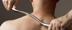
Instrument Assisted Soft Tissue Mobilization, also known as ”IASTM” for short is a process in which the clinician uses a set of ergonomically designed hand held instruments to break down the scar tissue and fascial restrictions in soft tissue (muscles, ligaments, tendons, and fascia ). The instruments you see in the photo are made of surgical grade stainless steel. Hypoallergenic aqueous cream or massage oil is used to facilitate gliding of the instrument along the muscle, tendon etc. during treatment.  The technique itself is said to have evolved from a form of Traditional Chinese Medicine called Gua Sha.  IASTM is a is a procedure that is growing  rapidly in popularity due to both the effectiveness and efficiency 0f the technique.

Once the damaged areas are detected, the instruments are used to deliver controlled microtrauma to the affected area. The purpose of the microtrauma is to stimulate a local inflammatory response, which initiates reabsorption of excessive scar tissue and facilitates a cascade of healing activities. Adhesions within the soft tissue that may have developed as a result of surgery, immobilization, muscle tears, or repetitive strain etc., are broken down, allowing the patient to regain function and range of movement. This treatment is a little intense, but it is extremely effective. It is somewhat similar to ”deep tissue massage” and/or ”rolfing”, but I always feel more is achieved in a shorter time-frame with IASTM technique . As in any Manual therapy treatment, it is usually not the only modality used. Supplementation with  stretching/strengthening exercises  etc. designed to correct biomechanical deficiencies by readdressing musculo-skeletal strength and imbalances  may also be prescribed  in conjunction with IASTM. I have used this form treatment both on myself and clients to great effect.

For more information check out these videos

Video 1

Video 2

Video 3


We are physiotherapists in Tralee, Co. Kerry. For more information on our treatments, prices, conditions we treat etc, check out our homepage. Also we are open 7am – 10pm weekdays and 8am – 2pm Saturdays. Phone 086-7700191