Snapping Hip
Illustration shows the bones of the hip joint, as well as the ligaments, tendons, and bursae that surround and protect the joint. Reproduced from The Body Almanac. © American Academy of Orthopaedic Surgeons, 2003.

Snapping Hip – Physiotherapist Tralee

Snapping hip syndrome is a clinical condition characterized by a palpable or audible snapping sensation in the hip area when walking, climbing a stairs, squatting or swinging your leg around etc. Although the condition is usually painless and harmless, the sensation can be annoying. In some cases, snapping hip leads to a bursitis, a painful swelling of the fluid-filled sacs that cushion tendons from bones of the hip joint, during movement. Snapping hip can also occur in other areas of the hip where tendons and muscles slide over knobbly areas on the hip bones. These areas include :

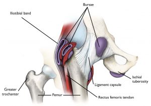
  • Outside of the hip : This is the most common,  where the iliotibial band passes over the portion of the thighbone known as the greater trochanter. When the hip is straight, the iliotibial band is behind the trochanter. When the hip bends, the band moves over the trochanter so that it is in front of it. The iliotibial band is always tight, like a stretched rubber band. Because the trochanter juts out slightly, the movement of the band across it creates the snap you hear. Eventually, snapping hip may lead to hip bursitis.
  • Front of the hip. Another tendon at the front of the hip that could cause snapping hip runs from the front of the thigh up to the pelvis  is the rectus femoris tendon.  As you bend the hip, the tendon shifts across the head of the thighbone, and when you straighten the hip, the tendon moves back to the side of the thighbone. This back-and-forth motion across the head of the thighbone causes the snapping.  The iliopsoas tendon can also catch on bony prominences at the front of the pelvis bone.
  • Back of the hip. Snapping in the back of the hip can involve the hamstring tendon. This tendon attaches to the sitting bone, called the ischial tuberosity. When it moves across the ischial tuberosity, the tendon may catch, causing a snapping sensation in the buttock region. This is rare.

Cause of snapping hip

It is most often the result of tightness in the muscles and tendons surrounding the hip. Athletes in sports  that require repetitive hip flexing are more likely to experience snapping hip. Young athletes can also be prone because tightness in the muscle structures of the hip due to adolescent growth spurts.


Treatment

Stretching the muscles of the affected areas ie. stretching of piriformis, tensor fascia latae, psoas/iliopsoas muscle, rectus femoris. Sometimes strengthening of the hip area may also be required to balance out the area, but usually the condition is a result of tight muscles and/or overuse, so stretching and rest, or activity modification is most effective.

In the rare instances that snapping hip does not respond to conservative treatment, your doctor may recommend surgery. The type of surgery will depend on the cause of the snapping hip. A bursitis can be cut out with surgery or often a cortisone injection is enough to settle it down.


Interesting articles on the condition include:

https://www.ncbi.nlm.nih.gov/pmc/articles/PMC4961351/

https://link.springer.com/article/10.1007/s00167-020-06305-w


For more information phone us and talk to one of our physiotherapists in Tralee, or click here.

Chondromalacia Patella – Knee Pain – Patellar Tracking Dysfunction

Chondromalacia patella
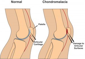
Chondromalacia patella, also known as ”Runners Knee” is one of the most common causes of knee pain in runners. The condition results from irritation of the cartilage on the under-surface of the kneecap. This cartilage is smooth and the kneecap normally glides effortlessly across it during bending of the knee joint. In some individuals however the kneecap does not track so smoothly due to poor alignment and the cartilage surface becomes irritated, resulting in inflammation and knee pain. In more severe cases there can be breakdown of the cartilage. Chondromalacia patella can affect athletes of any age but tends to be more common in women, most likely due to anatomical differences between the sexes ie. wider hips in females which results in a greater angulation between hip and knee, thus resulting in increased lateral forces on the patella.


Chondromalacia Patella – Causes

There are several causes both structural and dynamic which are linked to the condition. These include excessive foot pronation(feet turn out when running etc.), tight IT band, tight vastus lateralis(basically outer lower quad), weak or slow firing vastus medialis (basically lower inner quad), increased Q angle (simply put the angle between the outer hip and centre of the knee), a lateral femoral condyle that is not sufficiently prominent anteriorly (simply put the knee joint does not fit together properly),and a small or high riding patella(knee cap).(McConnell, 2002)


Chondromalacia Patella – Symptoms

The most common symptom is a dull, aching pain in the front of the knee, behind the kneecap. This pain is often worse when you go up or down stairs. It also can flare up after you have been sitting in one position for a long time. For example, your knee may be painful and stiff when you stand up after watching a movie or after a long trip in a car or plane. In some cases, the painful knee also can appear puffy or swollen. Chondromalacia can sometimes cause a creaky sound or grinding sensation known as ”crepitus” when you move your knee.


Chondromalacia Patella – Physio Treatment 

Suitable treatment may involve 1. Soft tissue work to loosen tightened structures such as vastus lateralis muscle, IT band, lateral retinaculum etc., 2.Strengthening of weak structures such as vastus medialis, glutes , hip abductors etc., 3. Correction of overpronation using orthotics, 4. Non steroidal anti-inflammatories such as ibuprofen to reduce pain and inflammation, 5. Rest with gradual return to exercise, 6. Taping to correct tracking can be a short term solution.(Hertling and Kessler, 2006) while you strengthen the vastus medialis muscle. Also there are supports you can purchase to help correct patella tracking while exercising. These are a good short term solution while you correct the problems referred to above. Here is a good example.

If nonsurgical treatments fail, or if you have severe symptoms, your doctor may recommend arthroscopy to check the cartilage inside your knee. If the cartilage is softened or shredded, damaged layers can be removed during the surgery, leaving healthy cartilage in place .


References

Hertling, D., Kessler, R.M. ”Management of Common Musculoskeletal Disorders : Physical Therapy Principles and Methods.” Lippincott, Philidelphia 524-533, 2006.

Mc Connell, J. ”The physical therapist’s approach to patellofemoral disorders.” Clinical Sports Medicine 21:363-387, 2002.


For more information on chondromalacia patella see this video.


If  you would like to get in touch to discuss your condition or make an appointment for physio in Tralee, please click

here for details.

Regards Eddie.

Supraspinatus tear
Fig. 1  Supraspinatus tear

A  Supraspinatus tear is a common area of injury in the rotator cuff complex. These are a group of muscles surrounding the humerus that help to keep the shoulder(humerous) in place. They are the supraspinatus, infraspinatus, subscapularis, and teres minor muscles. These muscles  facilitate movement of the shoulder joint in different directions, ie. raising your arm overhead. Other supportive structures which help to stabilise the shoulder include the shoulder capsule and the ligaments of the shoulder. See video.


Supraspinatus Tear – Acute vs Degenerative

Acute tears to the rotator cuff muscles/tendons often occur due to sports or impact injuries. can occur alongside injuries like shoulder dislocation, clavicle fractures, or other rotator cuff injuries that can happen as the result of things like a fall on your outstretched arm or attempting to lift something too heavy; plus there are a variety of sports where the athletes are prone to shoulder damage like baseball, basketball, rugby, AFL Football, and tennis. Construction work and other high-risk physical jobs can also increase the likelihood of experiencing this type of injury.

Alternatively degenerative type tears can occur due to overuse and as a result of age and lifestyle. Instead of a single catastrophic episode or trauma to the shoulder, these tears are the result of damage and wear on the supraspinatus structure slowly over time. The likeliness of these issues increases with age and is more common in the dominant hand. In addition, if you experience a degenerative tear in one shoulder, you’re at a greater risk for a tear in the other shoulder. Men over forty are the most likely to have degenerative supraspinatus tears. Factors like smoking, hypercholesterolemia, weight and BMI, height, bone spurs, and other genetic factors increase the chances as well.

A Supraspinatus tendon tear can be full thickness (the entire muscle is affected) or partial thickness (incomplete tear). Tears can be symptomatic , where there is pain and your ability to perform basic everyday tasks is greatly reduced,  or it can be asymptomatic, meaning that the tear  causes very slight or no pain.


Difference between full and partial thickness Supraspinatus tear

You can partially or fully tear your supraspinatus muscle, and remember that these sorts of tears can be symptomatic (meaning they cause supraspinatus pain and inhibit your range of motion and ability to perform everyday tasks) or asymptomatic, meaning the tear is present but it not currently causing you pain or otherwise causing problems in your life. Partial thickness tears of the supraspinatus muscle are an incomplete disruption of muscle fibers.

If you have been diagnosed with a partial thickness tear and begin experiencing more pain you should talk to your orthopaedic surgeon. Sometimes partial tears can progress to full thickness tears. Full thickness tears can also occur spontaneously and are the complete disruption of the fibers of the supraspinatus muscle.

The supraspinatus tendon can also be the site of injury. Tendons have poor blood supply and will not heal themselves. The longer these tears are left untreated, the more chance the tendon tear will enlarge and retract which results in more difficult surgery to repair this damage.


Diagnosis of a Supraspinatus tear

Various orthopedic tests can help indicate the likelihood of a supraspinatus tear during a physical exam. Magnetic resonance imaging (MRIs), and ultrasound scans are all used to confirm diagnosis.


Treatment

Depending on the severity of the tear, a surgeon may recommend starting with a non-surgical treatment like physiotherapy.  For most tears this is rarely effective. Cortisone injections can give short term relief but cause more harm long term. They can be effective if the main cause of pain is a sub-acromium bursitis

For full thickness tears and more major tears  there is significant damage to the tendon, various surgical procedures such as arthroscopic shoulder surgery are usually required. Full thickness tears will not heal without surgery. I have however seen cases where the supraspinatus muscle shrivels up after a full tear and basically withers away. In some such cases there is little or no pain at that stage and other muscles take over to a certain extent during shoulder movement, allowing the person to carry out normal daily activities.


To discuss your condition, make an appointment or get a second opinion please contact us.