Chondromalacia Patella – Knee Pain – Patellar Tracking Dysfunction

Chondromalacia patella
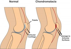
Chondromalacia patella, also known as ”Runners Knee” is one of the most common causes of knee pain in runners. The condition results from irritation of the cartilage on the under-surface of the kneecap. This cartilage is smooth and the kneecap normally glides effortlessly across it during bending of the knee joint. In some individuals however the kneecap does not track so smoothly due to poor alignment and the cartilage surface becomes irritated, resulting in inflammation and knee pain. In more severe cases there can be breakdown of the cartilage. Chondromalacia patella can affect athletes of any age but tends to be more common in women, most likely due to anatomical differences between the sexes ie. wider hips in females which results in a greater angulation between hip and knee, thus resulting in increased lateral forces on the patella.


Chondromalacia Patella – Causes

There are several causes both structural and dynamic which are linked to the condition. These include excessive foot pronation(feet turn out when running etc.), tight IT band, tight vastus lateralis(basically outer lower quad), weak or slow firing vastus medialis (basically lower inner quad), increased Q angle (simply put the angle between the outer hip and centre of the knee), a lateral femoral condyle that is not sufficiently prominent anteriorly (simply put the knee joint does not fit together properly),and a small or high riding patella(knee cap).(McConnell, 2002)


Chondromalacia Patella – Symptoms

The most common symptom is a dull, aching pain in the front of the knee, behind the kneecap. This pain is often worse when you go up or down stairs. It also can flare up after you have been sitting in one position for a long time. For example, your knee may be painful and stiff when you stand up after watching a movie or after a long trip in a car or plane. In some cases, the painful knee also can appear puffy or swollen. Chondromalacia can sometimes cause a creaky sound or grinding sensation known as ”crepitus” when you move your knee.


Chondromalacia Patella – Physio Treatment 

Suitable treatment may involve 1. Soft tissue work to loosen tightened structures such as vastus lateralis muscle, IT band, lateral retinaculum etc., 2.Strengthening of weak structures such as vastus medialis, glutes , hip abductors etc., 3. Correction of overpronation using orthotics, 4. Non steroidal anti-inflammatories such as ibuprofen to reduce pain and inflammation, 5. Rest with gradual return to exercise, 6. Taping to correct tracking can be a short term solution.(Hertling and Kessler, 2006) while you strengthen the vastus medialis muscle. Also there are supports you can purchase to help correct patella tracking while exercising. These are a good short term solution while you correct the problems referred to above. Here is a good example.

If nonsurgical treatments fail, or if you have severe symptoms, your doctor may recommend arthroscopy to check the cartilage inside your knee. If the cartilage is softened or shredded, damaged layers can be removed during the surgery, leaving healthy cartilage in place .


References

Hertling, D., Kessler, R.M. ”Management of Common Musculoskeletal Disorders : Physical Therapy Principles and Methods.” Lippincott, Philidelphia 524-533, 2006.

Mc Connell, J. ”The physical therapist’s approach to patellofemoral disorders.” Clinical Sports Medicine 21:363-387, 2002.


For more information on chondromalacia patella see this video.


If  you would like to get in touch to discuss your condition or make an appointment for physio in Tralee, please click

here for details.

Regards Eddie.

Achilles tendon ruptures will debilitate your lower leg completely, the moment they happen. The Achilles tendon is an important part of the leg. It is located just behind and above the heel, attaching the heel bone to the calf muscles. Its function is to help in bending the foot downwards at the ankle (this movement is called plantar flexion). If the Achilles tendon is torn/ruptured, the tear may be either partial or complete. In a partial tear, the tendon is torn but is still partly joined to the calf muscle. With complete tears, the tendon is completely torn and the connection between the calf muscles and the ankle bone is lost. If your Achilles tendon is ruptured you will be unable to stand on your tiptoes and you will have a flat-footed walk with a severe limp, along with considerable pain initially.


An Achilles tendon can tear when there is too high a load or stress placed on it. This can happen with activities involving a forceful push off with the foot – ie. running/sprinting in football, basketball, tennis etc. I have also seen it occur when somebody sprints off suddenly on a sandy beach, the sand giving way under the foot putting extra stress on the calf and the achilles tendon. The push off movement uses a strong contraction of the calf muscles maximally stressing both the calf muscles and the Achilles tendon . Injury to the achilles can also occur due to falls, if the foot is suddenly forced into an upward-pointing position, stretching the tendon. Another possible injury is a deep cut/laceration to the tendon.


Achilles Tendon Rupture
   Fig. 1.  Achilles Tendon Rupture


Sometimes the Achilles tendon is weak, making it more prone to rupture. Factors that weaken the Achilles tendon are:

  • Corticosteroid medications (such as prednisolone) – mainly when used as long-term treatment.
  • A corticosteroid injection near the Achilles tendon.
  • Certain rare medical conditions, such as Cushing’s syndrome, where the body makes too much of its own corticosteroid hormones.
  • Tendinopathies of the Achilles tendon.
  • Other medical conditions which can make the tendon more prone to rupture; for example, rheumatoid arthritis, diabetes, gout and systemic lupus erythematosus.
  • Certain antibiotic medicines may slightly increase the risk of having an Achilles tendon rupture. These are the quinolone antibiotics such as ciprofloxacin and ofloacin. The risk of having an Achilles tendon rupture with these antibiotics is quite low and mainly applies to people who are also taking corticosteroid medication.


You might hear a snap or feel a sudden sharp pain when the tendon is torn (ruptured)during a sporting activity or injury. The sharp pain usually settles quickly, although there may be some aching at the back of the lower leg. After the injury, the usual symptoms are:

  • A flat-footed type of walk. You can walk and bear weight but cannot push off the ground properly on the side where the tendon is ruptured.
  • Inability to stand on tiptoe.
  • If the tendon is completely torn, you may feel a gap just above the back of the heel. However, if there is bruising and swelling may disguise the gap.

An Achilles tendon rupture is usually diagnosed based on symptoms, history of the injury and a physio/doctor’s examination. An orthopedic test called Thompson’s test (also known as the calf squeeze test) may help diagnosis. In this test, you will be asked to lie face down on the examination bench and to bend your knee. The doctor will gently squeeze the calf muscles at the back of your leg and observe how the ankle moves. If the Achilles tendon is OK, the calf squeeze will make the foot point away from the leg (a movement called plantar flexion). This is quite an accurate test for Achilles tendon ruptures. An ultrasound or MRI may be used to confirm diagnosis and give a fuller picture of the injury.


Treatment and recovery

Treatment for a ruptured achilles tendon usually involves surgery to repair the tendon. The surgeon sews/sticks together the torn ends of the Achilles tendon, and may also use another tendon or a tendon graft to help with the repair. A plaster cast or brace is needed after the operation for about eight weeks, to keep the foot immobilized, allowing the tendon can heal. The plaster cast or the brace is positioned so that the foot is pointing slightly downwards, which takes the strain off the tendon.

Traditionally, crutches were used to keep weight off the leg during the first few weeks of treatment. Current thinking tends towards using the leg normally early on (early mobilization). This involves fitting a plaster cast or a brace which you can walk on. It is more convenient because you do not need to use crutches. Physiotherapy will also be needed, especially when the cast is removed. I have personally found instrument assisted soft tissue work(after about 8 weeks) to be very beneficial along with stretching/strengthening work and deep tissue work to the calf during recovery. Full recovery is greatly slowed when there is no hands-on work done during recovery, after the cast is removed once an ok is given by the surgeon to commence.

Depending on a person’s profession, some people may need several weeks off work after an Achilles tendon tear (rupture); the time taken to return to sport is usually between 4 and 12 months. Generally, the outlook is good. However, the tendon does take time to heal, usually about six to eight weeks. More time will be needed after this to allow the muscles and tendon to regain normal strength.


For more on rehabilitation on achilles tendon ruptures post surgery click here.


Physiotherapist in Tralee. Ring 08677001 to discuss your condition, book an appointment or get a second opinion.

stenosis of spine
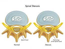
Spinal stenosis is a narrowing of the spaces within your spine, which can put pressure on the nerves that travel through the spine.  Others may experience pain, tingling, numbness and muscle weakness. Symptoms often start gradually and worsen over time. Spinal stenosis is most commonly caused by wear-and-tear changes in the spine related to osteoarthritis. In severe cases of spinal stenosis, doctors may recommend surgery to create additional space for the spinal cord or nerves. Most people with spinal stenosis are over the age of 50. There are two main types of spinal stenosis: 1. Cervical stenosis. where the narrowing occurs in  your neck. 2. Lumbar stenosis where the narrowing occurs in the lower back.


Symptoms of spinal stenosis

Neck (cervical spine)

  • Numbness or tingling in a hand, arm, foot or leg
  • Weakness in a hand, arm, foot or leg
  • Problems with walking and balance
  • Neck pain
  • In severe cases, bowel or bladder dysfunction (urinary urgency and incontinence)

Lower back (lumbar spine)

  • Numbness or tingling in a foot or leg
  • Weakness in a foot or leg
  • Pain or cramping in one or both legs when you stand for long periods of time or when you walk, which usually eases when you bend forward or sit
  • Back pain

Causes of spinal stenosis

The backbone (spine) runs from your neck to your lower back. The bones of your spine form a spinal canal, which protects your spinal cord (nerves). Some people are born with a small spinal canal. But most spinal stenosis occurs when something happens to narrow the open space within the spine, such as the following :

  • Overgrowth of bone. Wear and tear damage from osteoarthritis on your spinal bones can prompt the formation of bone spurs, which can grow into the spinal canal.
  • Herniated disks. The soft cushions that act as shock absorbers between your vertebrae tend to dry out with age. Cracks in a disk’s exterior may allow some of the soft inner material to escape and press on the spinal cord or nerves.
  • Thickened ligaments. The tough cords that help hold the bones of your spine together can become stiff and thickened over time. These thickened ligaments can bulge into the spinal canal.
  • Tumors. Abnormal growths can form inside the spinal cord, within the membranes that cover the spinal cord or in the space between the spinal cord and vertebrae. These are uncommon and identifiable on spine imaging with an MRI or CT.
  • Spinal injuries. Car accidents and other trauma can cause dislocations or fractures of one or more vertebrae. Displaced bone from a spinal fracture may damage the contents of the spinal canal. Swelling of nearby tissue immediately after back surgery also can put pressure on the spinal cord or nerves.


In severe cases of spinal stenosis, doctors may recommend surgery to create additional space for the spinal cord or nerves.


See video below for some tips.

https://youtu.be/GbJgE0LPKCk


Physio in Tralee specializing in the treatment of back pain, neck pain, foot pain and sports injuries.