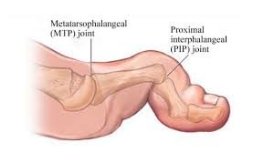
What is Hallux Rigidus?

Hallux Rigidus

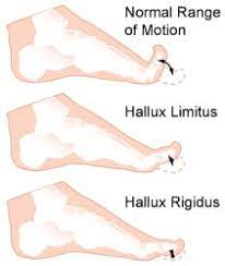
Big toe joint arthritis (otherwise known as Hallux Rigidus) is a form of degenerative arthritis. In this condition, surfaces of the joints in the big toe begin to wear away and extra bone can also develop in the form of  bone spurs or osteophytes. This limits the movement of the joint. The big toe  needs to bend significantly when stepping off. Consequently, arthritis in this joint can greatly affect walking, running etc.

Hallux Rigidus is a condition that tends to get worse over time. In it’s earlier stages, it may be referred to as Hallux Limitus, which is where there is limited movement of the big toe joint. With progression, Hallux Rigidus develops which can result in stiffness developing in the big toe joint and there may also be swelling.

To compensate for pain in the big toe joint, people with Hallux Rigidus tend to adjust their walking pattern which can also result in knee, hip or lower back pain. There is often difficulty finding shoes that fit properly shoes due to pain and/or inflammation in the joint. Early diagnosis can be made by physical examination and x-ray. Early treatment gives the best chance of avoiding surgery.

What causes Hallux Rigidus?

The big toe joint is designed to bear a considerable amount of stress during walking. However faulty biomechanics or structural abnormalities such as flat feet can stop the big toe from bending normally, and eventually lead to the development of osteoarthritis in the big toe joint. Other factors that can increase the risk of developing this condition include genetics (eg, having a certain foot type), injury to the big toe , other inflammatory diseases, and working in a job that places excessive stress on the big toe joint .

Treatment

In cases of Hallux Rigidus, certain types of footwear and orthotics may be recommended to reduce pressure and motion on the big toe.  Custom orthotics that conform very closely to the arch of the foot are most effective in improving big toe joint function. These orthotics may incorporate  something like a 3mm kinetic wedge in combination with a built in forefoot posting to take the pressure off the big toe joint. Ice and anti-inflammatory medication can also be used to reduce inflammation and pain. If it does not respond to conservative treatment, surgery may be recommended as a last resort in more severe cases.

Posterior ankle impingement physiotherapy
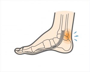
Posterior ankle impingement is a condition characterised by tissue damage at the back of
the ankle joint due to compression of these structures. This occurs when the foot and ankle
are pointed maximally away from the body (plantarflexion – figure 1. ). It may occur when
compressive forces are too repetitive and/or too forceful. This can occurs in the presence of
ankle swelling or bony anomalies, such as additional bone, a condition known as an “os
trigonum”. Posterior ankle impingement is most commonly found in gymnasts, ballet
dancers, and footballers, because they regularly maximally plantarflex their ankles during
their activities. The condition can also occur due to inadequate rehabilitation of an acute
ankle injury (ie. ankle sprain).

Mechanism of Injury

Posterior ankle impingement may develop due to an acute traumatic plantar hyperflexion
event, such as an ankle sprain. It may also occur as a result of repetitive low-grade trauma
associated with plantar hyperflexion, say like in case of a female ballet dancer. It is
important to differentiate between these two, because the latter, that is posterior
impingement from overuse, has a better prognosis.
The anatomy of the posterior ankle is a key factor in the occurrence of posterior
impingement syndrome . The more common causes of the condition are osseous in nature,
such as the os trigonum, an elongated posterolateral tubercle of the talus (known as
Stieda’s process), a downward sloping posterior lip of the tibia, an osteophyte from the
posterior distal tibia , or a prominent posterior process of the calcaneus. However, posterior
impingement can also be soft tissue related, as with a thickened posterior joint capsule ,
post-traumatic scar tissue, post-traumatic calcifications of the posterior joint capsule, or
loose bodies in the posterior part of the ankle joint. Symptoms for all of these conditions
relate to physical impingement of osseous or soft tissue structures, resulting in painful
limitation of the full range of ankle movement.
The most common cause ''os trigonum'' is an extra (accessory) bone that sometimes
develops behind the ankle bone (talus). The mineralized os trigonum appears between the
ages of 7 and 13 years and usually fuses with the talus within 1 year, forming the trigonal
(Stieda) process. It may remain as a separate ossicle in 7-14% of patients, and is often
bilateral(in both ankles). An os trigonum can be a focus of osseous abutment against other
structures. Pain can also be caused by disruption of the cartilaginous synchondrosis
between the os trigonum and the lateral talar tubercle as a result of repetitive microtrauma
and chronic inflammation.
In the case of soft tissue impingement it usually results from scarring and fibrosis associated
with synovial, capsular, or ligamentous injury ie. bad ankle sprain. It is thought that this
type of manifestation usually usually occurs when a significant soft-tissue component
forms. The soft-tissue component can consist of synovial thickening throughout the
posterior capsule or be more focal, involving the posterior intermalleolar or talofibular ligament. The flexor hallucis longus tendon runs in the groove between the lateral and
medial processes of the talus and can also be injured in posterior impingement, resulting in
tenosynovitis.


Signs and symptoms

Patients who have posterior impingement complain of chronic deep posterior ankle pain
worsened by forced plantar flexion or push-off forces as occur during activities such as
ballet dancing, jumping, or running downhill. In some patients, forced dorsiflexion(opposite
to plantarflexion) is also painful. Physical examination reveals pain on palpation over the posterolateral talar process, which is located along the posterolateral aspect of the ankle between the Achilles and peroneal
tendons . Passive forced plantar flexion results in pain and often a grinding
sensation as the posterolateral talar process is entrapped between the posterior tibia and
calcaneus.


Diagnosis of posterior ankle impingement

A thorough examination by an experienced practitioner may be all that is necessary to
diagnose posterior ankle impingement. Further investigations such as an X-ray, MRI, CT scan
or Ultrasound may help confirm diagnosis.


Physiotherapist in Tralee, Co. Kerry………..Phone 0867700191 to make an appointment or discuss your condition.

Temperomandibular Joint Disorder
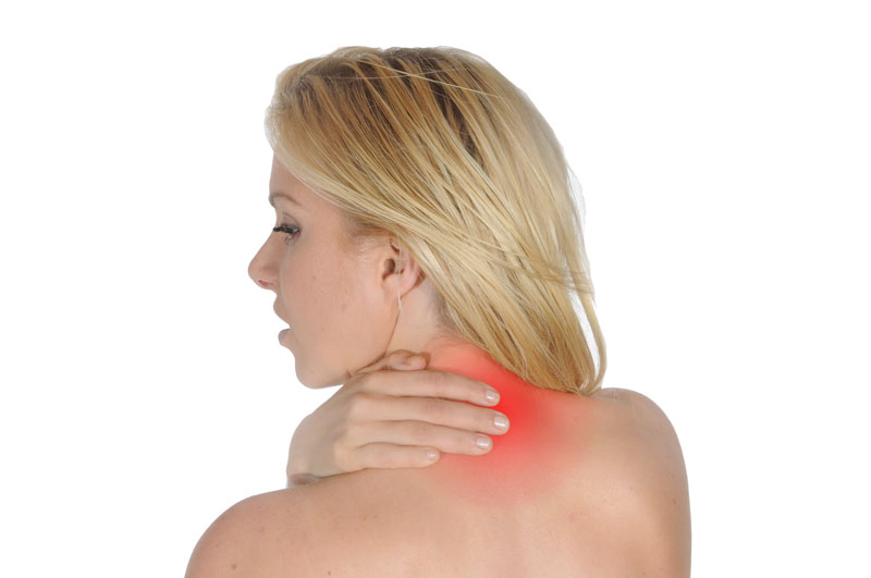
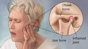
The temporomandibular joint works as a combination of hinge and sliding actions. It connects the jawbone to the skull on each side of the face.  The parts of the bones that interact in the joint are covered with cartilage and are separated by a small shock-absorbing disk, which normally keeps the movement smooth.  Temporomandibular  joint disorder (TMJD) can cause pain in your jaw joint and in the muscles that control jaw movement.

Symptoms

  • Pain in one or both of the temporomandibular joints
  • Difficulty or pain chewing
  • Locking of the joint, making it difficult to open or close your mouth
  • Clicking sound or grating sensation when you open your mouth or chew

Causes of  Temporomandibular Joint Disorder

Painful TMJ disorders can occur if:

  • The disk erodes or moves out of its proper alignment
  • The joint’s cartilage is damaged by arthritis
  • The joint is damaged by a blow or other impact

In many cases, however, the cause of TMJ disorders is unclear.

Treatment of  Temporomandibular Joint Disorder

In some cases, the symptoms of TMJ disorders may go away without treatment. Some of the following may also help.

Medications

  • Pain relievers and anti-inflammatories. These can help relieve pain and inflammation.
  • Muscle relaxants. These can help relax the jaw muscles.

Therapies

Nondrug therapies for TMJ disorders include:

  • Oral splints or mouth guards.  These devices worn at night while sleeping can help prevent grinding of the teeth.
  • Physical therapy.  Deep tissue massage work on neck and jaw muscles can be beneficial along with certain rehabilitation exercises.
  • Education. Education can help you understand the factors and behaviors that may aggravate your pain, so you can avoid them. Examples include teeth clenching or grinding, eating foods where you have to open the jaw wide like apples and burgers.

When conservative treatments fail, the following may be considered:

  • Arthrocentesis. This is a minimally invasive procedure that involves the insertion of small needles into the joint so that fluid can be irrigated through the joint to remove debris and inflammatory byproducts.
  • Injections. In some people, corticosteroid injections into the joint may be helpful. Infrequently, injecting botulinum toxin type A (Botox, others) into the jaw muscles used for chewing may relieve pain associated with TMJ disorders.
  • TMJ arthroscopy.  A small thin tube is placed into the joint space, an arthroscope is then inserted and small surgical instruments are used for surgery. TMJ arthroscopy has fewer risks and complications than open-joint surgery does, but it has some limitations as well.
  • Modified condylotomy. Modified condylotomy addresses the TMJ indirectly, with surgery on the mandible, but not in the joint itself. It may be helpful for treatment of pain and if locking is experienced.
  • Open-joint surgery. If your jaw pain does not resolve with more-conservative treatments and it appears to be caused by a structural problem in the joint, your doctor or dentist may suggest open-joint surgery (arthrotomy) to repair or replace the joint. However, open-joint surgery involves more risks than other procedures do.


For some handy self treatment tips, check out this video https://youtu.be/7b73yE0U2t0

Tmj manipulation video


Check out our website and feel free to contact us to discuss your condition or to set up an appointment.


When Back Pain Is A Symptom Rather Than A Condition

Back pain may sometimes be a symptom rather than a condition as such. A practitioner needs to always keep this at the back of their mind when treating or assessing somebody. Sometimes things need more investigation. A good physio will spot this early and refer you on quickly to your G.P. for further examination. It is always better to play it safe if in doubt.  I always believe you should be seeing improvement in your condition from treatment to treatment. If there is no improvement after several treatments you need to be asking yourself a few questions i.e. Do I need to try a different physio or do I need to consult my doctor for a second opinion. Sometimes the proper treatment protocol for a condition is all that is needed for a swift recovery. The list below is not meant to be alarmist. It is more to emphasize the fact that continuous unrelenting back pain can be a symptom of something else going on within your body.


Below are examples of conditions/medications etc. that may have back pain as a side effect/symptom.


kidney stones – A kidney stone may not cause symptoms until it moves around within your kidney or passes into your ureter (the tube connecting the kidney and bladder). At that point, you may experience symptoms such as severe pain in your side and back below the ribs, or pain that radiates into the lower abdomen and groin.

Lupus is a long-term autoimmune disease in which the body’s immune system becomes hyperactive and attacks normal, healthy tissue. Symptoms include inflammation, swelling, and damage to the joints, skin, kidneys, blood, heart, and lungs. Lupus can cause neck and back pain, because muscles in these areas can become inflamed due to the lupus. Furthermore, the muscle pain syndrome  ”fibromyalgia”  can cause pain in these areas and is commonly associated with lupus.

Spinal arthritis/facet joint arthritis causes stiffness and back pain.

Cancers – A primary bone cancer tumor in the spine can cause back pain, as can a number of other cancers when they have metastasized(spread to other sites in the body), such as breast cancer, testicular cancer, colon cancer, and lung cancer. In fact, back pain is often the one of first symptoms that people with lung cancer notice before they are diagnosed. A tumor in the lungs can put pressure on the spine, or can affect the nerves around the chest wall and spine.

Spondylosis a painful condition of the spine resulting from the degeneration of the intervertebral discs.

Spondylitis is a condition resulting in inflammation within the joints of the spine. As the inflammation goes and healing takes place, bone grows out from both sides of the vertebrae and may join the two together, causing a stiffening known as ankylosis. The progressed condition is called ankylosing spondylitis. The cause is not yet known.

back pain


Spondylolisthesis is a slipping of vertebra that occurs, in most cases, at the base of the spine.

Spondylolysis is a defect or fracture of one or both wing-shaped parts of a vertebra, can result in vertebrae slipping backward, forward, or over a bone below.


Spondylosis, Spondylitis, Spondylolisthesis, Spondylolysis all have back pain as a symptom.


Fractures – Even a minor fracture along the spine  can cause considerable back pain.

StatinsStatins are drugs that can help lower your cholesterol. One of the more severe side effect of statins is myotoxicity(having a toxic effect on muscle), in the form of myopathy(a disease of the muscle in which the muscle fibers do not function properly. This results in muscular weakness), myalgia(muscle pain), myositis(inflammation and degeneration of muscle tissue) or rhabdomyolysis(a condition in which damaged skeletal muscle breaks down). Currently, the only effective treatment of statin-induced myopathy is the discontinuation of statin use in patients affected by muscle aches, pains and elevated creatine kinase levels. Creatine kinase are the clinical measure of muscle damage (rhabdomyolysis).

Anticonvulsant drugs such those used in the treatment of conditions like epilepsy can cause changes in calcium and bone metabolism. This may in time lead to decreased bone mass and a risk of osteoporotic fractures in the spine which may also result in severe back pain. Two widely used antiepileptic drugs phenytoin and carbamazepine are recognized to have direct effects on bone cells.

Corticosteroids – Longterm use of corticosteroids increase the risk of compression fractures in the spine(back).


Neurological Compromise — A Red Flag

Neural compromise can result from spinal cord or cauda equina compression . Cauda equina compression usually results from a fracture, tumor, epidural hematoma, or abscess, and occasionally from a massive disk herniation. Paraplegia(impairment in motor or sensory function of the lower extremities), quadriplegia(paralysis caused by illness or injury that results in the partial or total loss of use of all four limbs and torso), or cauda equina deficit should trigger an aggressive search for the cause.

Cauda equina compression classically presents with back pain, bilateral sciatica(pain down the back of both legs), saddle anesthesia, and lower extremity weakness progressing to paraplegia, but in practice these symptoms are variably present and diagnosing the condition often requires a high degree of suspicion. Hyporeflexia(no reflexes) is typically a sign of cauda equina compression, while hyperreflexia(overresponsive reflexes), clonus(series of involuntary, rhythmic, muscular contractions and relaxations), and the Babinski sign(see below *)  suggest spinal cord compression, requiring an evaluation of the cervical and thoracic spine. Cauda equina compression typically involves urinary retention; in contrast, cord compression typically causes incontinence. If either cauda equina or spinal cord compression is detected during an initial examination, an immediate more extensive evaluation is warranted. MRI is the study of choice.

*(The Babinski reflex occurs after the sole of the foot has been firmly stroked. The big toe then moves upward or toward the top surface of the foot. The other toes fan out. This reflex is normal in children up to 2 years old. It disappears as the child gets older. It may disappear as early as 12 months. When the Babinski reflex is present in a child older than 2 years or in an adult, it is often a sign of a central nervous system disorder. The central nervous system includes the brain and spinal cord.)

Spinal epidural hematoma

Spinal epidural hematoma is a rare but dramatic cause of paralysis in elderly patients. In most cases, there is no antecedent trauma. Lawton et al. (1995), in a series of 30 patients treated surgically for spinal epidural hematoma, found that 73% resulted from spine surgery, epidural catheterization, or anticoagulation therapy. Other possible causes of epidural hematoma include vascular malformations, angiomas, aneurysms, hypertension, and aspirin therapy. The same study found that the time from the first symptom to maximal neurologic deficit ranged from a few minutes to 4 days, with the average interval being nearly 13 hours.

Although painless onset has been reported, spinal epidural hematoma typically presents with acute pain at the level of the lesion, which is often rapidly followed by paraplegia(impairment in motor or sensory function of the lower extremities) or quadriplegia(is paralysis caused by illness or injury that results in the partial or total loss of use of all four limbs and torso), depending on the location of the hemorrhage. Sometimes the onset of pain is preceded by a sudden increase of venous pressure from coughing, sneezing, or straining at stool. Urinary retention often develops at an early stage.

Most lesions occur in the thoracic region(rib area of back) and extend into the cervicothoracic(upper back and neck) or the thoracolumbar(ribs to lower back area) area. The pain distribution may be radicular(affecting or relating to the root of a spinal nerve), mimicking a ruptured intervertebral disk.

Evaluation should be with MRI. Early recognition, MRI confirmation, and treatment should be accomplished as soon as possible. Recovery depends on the severity of the neurologic deficit and the duration of symptoms before treatment. Lawton et al.(1995), found that patients taken to surgery within 12 hours had better neurologic outcomes than patients with identical preoperative neurologic status whose surgery was delayed beyond 12 hours. Surgery should not be withheld because of advanced age or poor health: in 10 reported cases in which surgery was delayed, all patients died.


For more about treatments and opening times etc. click on our homepage

bicipital-tendonitis
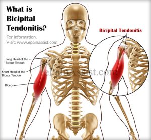
Bicipital tendonitis is a common cause of shoulder pain, often developing in people who perform repetitive, overhead movements. Biceps tendinitis develops over time, the pain being located at the front of the shoulder. The biceps muscle has two parts referred to as the long head and the short head. The tendon of the long head of the biceps is most commonly implicated with tendonitis. When this tendon is subjected to repetitive stresses, it can become irritated, swollen, and painful.This occurs where the tendon sits within the bicipital groove at the top of the humerus under the transverse ligament before it becomes part of the shoulder joint capsule.

Pain at this exact spot when pressed with a finger as the arm is rotated in and out while standing, is usually a fairly reliable test to confirm this condition. Imaging techniques such as MRI are typically not needed to diagnose biceps tendonitis.

Symptoms – Bicipital Tendonitis

Pain or tenderness in the front of the shoulder, which worsens with overhead lifting or activity.

Pain that moves down along the upper arm

An occasional snapping sound or sensation in the shoulder

Treatment – Bicipital Tendonitis

The initial goals of treatment for bicipital tendonitis are to reduce inflammation and swelling. Patients should restrict above shoulder height movements, reaching out with the affected arm and lifting. They should apply ice to the affected area for 10-15 minutes, 2-3 times daily for several days. Non-steroidal anti-inflammatory drugs (NSAIDs), such as ibuprofen, may help recovery. Probably the biggest aid to recovery is rest from the aggravating activity for several weeks. I often come across this condition in weight trainers who front press or incline bench press, the bar being too far out from their neck during pressing. It is also common in swimmers with poor technique or who ramp up their training distance/pace too quickly.


Physiotherapist Tralee : Phone 086-7700191 for an appointment, second opinion or to discuss your injury.

 Hammer Toes

Hammer toes and mallet toes are foot deformities that occur due to an imbalance in the muscles, tendons or ligaments that normally hold the toe straight. This is a deformity that causes your toe to bend or curl downward instead of pointing forward. It can affect any toe.

Hammer ToesHammer Toes

Causes of Hammer Toes

Hammertoe and mallet toe have been linked to:

  • An imbalance in toe muscles leading to instability, which in turn can cause the toe to contract.
  • Genetics(say one of your parents has it). It may be present at birth.
  • It may develop over time due to arthritis. Diabetes could also increase your risk.
  • An injury in which you stub, jam or break a toe can make it more likely for that digit to develop hammertoe.
  • Poorly fitting and/or high-heeled shoes or footwear that are too tight at the toes crowding your toes into a space in which they can’t lie flat.
  • toe length(if your second toe is too long)
  • Sex (women seem more prone but maybe that is down to fashion ie. high heels etc.?)

Complications of Hammer Toes

At first, a hammertoe or mallet toe might maintain its flexibility. Eventually, the tendons of the toe can contract and tighten, causing your toe to become permanently bent. The toe may become painful and moving it may also be difficult or painful. The raised portion of the toe or toes can rub against your shoes, causing painful corns or calluses.

Prevention of Hammer Toes

Relieving the pain and pressure of hammertoe may involve changing your footwear and/or wearing orthotics. If you have a more severe case of hammertoe or mallet toe, you might need surgery to get relief.

You can avoid many foot, heel and ankle problems with shoes that fit properly. Here’s what to look for when buying shoes:

  • Make sure there is adequate toe room. Avoid shoes with pointed toes.
  • Low heels. Avoiding high heels.
  • Make sure shoes are supportive and comfortable. Lace them up properly

These additional tips can help you buy the right shoes:

  • Buy shoes at the end of the day. Your feet swell slightly throughout the day.
  • Check your size. As you age, your shoe size might change — especially the width. Measure both feet and buy for the larger foot.
  • Buy shoes that fit. Be sure shoes are comfortable before you buy them. If necessary, a shoe repair store might be able to stretch shoes in tight spots, but it’s better to buy them to fit.

For more on hammer toes  see this video

Physiotherapy clinics in Tralee and Dingle phone Eddie on 086-7700191

Trigger Finger

trigger finger
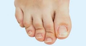
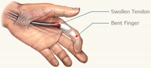
Trigger finger is a condition in which one of your fingers gets stuck in a bent position. Your finger may bend or straighten with a snap — like a trigger being pulled and released.

Tendons are fibrous cords that attach muscle to bone. Each tendon is surrounded by a protective sheath. Trigger finger occurs when the affected finger’s tendon sheath becomes irritated and inflamed. This interferes with the normal gliding motion of the tendon through the sheath. Prolonged irritation of the tendon sheath can produce scarring, thickening and the formation of bumps (nodules) in the tendon that impede the tendon’s motion even more.

People with work or hobbies that require repetitive gripping actions are at higher risk of developing trigger finger. The condition is also more common in women and people who suffer with diabetes.

Symptoms

Signs and symptoms of trigger finger may progress from mild to severe and include:

  • Finger stiffness, particularly in the morning
  • A popping or clicking sensation as you move your finger
  • Tenderness or a bump (nodule) on palmside at the base of the affected finger
  • Finger catching or locking in a bent position, which suddenly pops straight
  • Finger locked in a bent position, which you are unable to straighten

Trigger finger can affect any finger and triggering is usually more pronounced in the morning.


Diagnosis

Diagnosis of trigger finger doesn’t require any elaborate testing. Diagnosis is based on  medical history and a physical exam. During the physical exam, your doctor/physo will ask you to open and close your hand, checking for areas of pain, smoothness of motion and evidence of locking.  He should also feel your palm to see if there is a lump present. If the lump is associated with trigger finger, the lump will move as the finger moves because the lump is an area of swelling in part of the tendon that moves the finger.

Treatment

Trigger finger treatment varies depending on the severity and duration of the condition. Nonsteroidal anti-inflammatory drugs  may relieve the pain but are unlikely to relieve the swelling constricting the tendon sheath or trapping the tendon.

Conservative non-invasive treatments may include:

  • Rest. Avoid activities that require repetitive gripping, repeated grasping or the prolonged use of vibrating hand-held machinery until your symptoms improve. If you can’t avoid these activities altogether, padded gloves may offer some protection.
  • A splint. Your doctor may have you wear a splint at night to keep the affected finger in an extended position for up to six weeks. The splint helps rest the tendon.
  • Stretching exercises. Your doctor may also suggest gentle exercises to help maintain mobility in your finger.

If conservative treatment fails here are the other options.

  • Steroid injection. Injection of a steroid medication near to or into the tendon sheath may reduce inflammation and allow the tendon to glide freely again. This is the most common treatment, and it’s usually effective for a year or more in most people treated. Sometimes it takes more than one injection. For people with diabetes, steroid injections tend to be less effective.
  • Percutaneous release. After numbing your palm/finger, your doctor inserts a sturdy needle into the tissue around your affected tendon. Moving the needle and your finger helps break apart the constriction that is blocking the smooth motion of the tendon.This treatment may be done under ultrasound control, so the doctor can see where the tip of the needle is under the skin and to be sure it opens the tendon sheath without damaging the tendon or nearby nerves.
  • Surgery. Working through a small incision near the base of your affected finger, a surgeon can cut open the constricted section of tendon sheath. This is a last resort.


Update

Here is a video of exercises to get rid of trigger finger, I came across by the internet famous physical therapists Bob and Brad. These exercises have actually proved to be extremely effective  …….See video


Physio in Tralee phone 086-7700191

IASTM
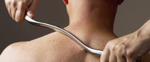
Instrument Assisted Soft Tissue Mobilization, also known as ”IASTM” for short is a process in which the clinician uses a set of ergonomically designed hand held instruments to break down the scar tissue and fascial restrictions in soft tissue (muscles, ligaments, tendons, and fascia ). The instruments you see in the photo are made of surgical grade stainless steel. Hypoallergenic aqueous cream or massage oil is used to facilitate gliding of the instrument along the muscle, tendon etc. during treatment.  The technique itself is said to have evolved from a form of Traditional Chinese Medicine called Gua Sha.  IASTM is a is a procedure that is growing  rapidly in popularity due to both the effectiveness and efficiency 0f the technique.

Once the damaged areas are detected, the instruments are used to deliver controlled microtrauma to the affected area. The purpose of the microtrauma is to stimulate a local inflammatory response, which initiates reabsorption of excessive scar tissue and facilitates a cascade of healing activities. Adhesions within the soft tissue that may have developed as a result of surgery, immobilization, muscle tears, or repetitive strain etc., are broken down, allowing the patient to regain function and range of movement. This treatment is a little intense, but it is extremely effective. It is somewhat similar to ”deep tissue massage” and/or ”rolfing”, but I always feel more is achieved in a shorter time-frame with IASTM technique . As in any Manual therapy treatment, it is usually not the only modality used. Supplementation with  stretching/strengthening exercises  etc. designed to correct biomechanical deficiencies by readdressing musculo-skeletal strength and imbalances  may also be prescribed  in conjunction with IASTM. I have used this form treatment both on myself and clients to great effect.

For more information check out these videos

Video 1

Video 2

Video 3


We are physiotherapists in Tralee, Co. Kerry. For more information on our treatments, prices, conditions we treat etc, check out our homepage. Also we are open 7am – 10pm weekdays and 8am – 2pm Saturdays. Phone 086-7700191


”Shin Splints” – Which Type Have You ?

shin splints”Shin splints” is a catch-all term for shin pain either on the front outside part of the lower leg (anterior shin splints) or on the inside of the lower leg (medial shin splints). It is the curse of many athletes including runners, tennis players, dancers etc. Frequently the condition plagues novice runners who do not build their mileage gradually enough. It also affects seasoned runners who abruptly change their workout regimen, suddenly adding too much mileage, or switching from running on the flat to hills. The term mainly refers mainly to the following three conditions 1. mini stress fractures within the tibia bone, 2. chronic exertional compartment syndrome, 3. medial tibial stress syndrome.  It is important to differentiate between the three for treatment.


1. Real ”shin splints”

Real ”shin splints” are mini stress fractures (splint-ers) within the tibia bone. With this condition pain is gradual in onset, getting worse with activity, and there is usually a history of an increase in training intensity. Pain may occur with walking, at rest, or even at night in bed. Treatment for this condition involves rest for about eight weeks from running to allow the little stress fractures to heal. You should be able to keep up fitness levels by cycling, swimming etc., as these exercises are low impact. You may need to look at lower limb biomechanics, running style, training practices etc. to prevent recurrence of the shin splints .


2. Shin splints – Chronic exertional compartment syndrome

Chronic exertional compartment syndrome is defined as increased pressure within a closed fibro-osseous space(like the space the tibia and fibula), causing reduced blood flow and tissue perfusion(perfusion is the process of a body delivering blood to a capillary bed in its biological tissue), which subsequently leads to ischemic pain(pain due to restriction of blood supply, and thus oxygen and nutrients to tissue) and possible permanent damage to tissues of the compartment. The syndrome is frequently bilateral (both legs). Typical features of the condition are absence of pain at rest, with increasingly achy pain and a sensation of tightness in the shins upon exertion. Symptoms usually resolve or significantly dissipate within several minutes of resting. Anyone can develop the condition, but it is more common in athletes who participate in activities that involve repetitive impact, such as running. Sometimes Chronic exertional compartment syndrome may respond to deep tissue work and myofascial release of the structures involved. Changing your chosen activity to one involving less impact may also help. Surgery may be used as a last resort to relieve the pressure. It involves operating on the inelastic tissue encasing each muscle compartment (fascia). Methods include either cutting open the fascia of each affected compartment (fasciotomy) or actually removing part of the fascia (fasciectomy).


Shin splints – Medial tibial stress syndrome

 Medial tibial stress syndrome is an inflammation of the muscles, tendons, and bone tissue around your tibia. A common cause of Medial Tibial Stress Syndrome is pes planus (flat feet) or over-pronation of the foot during running. This puts increased strain on the Tibialis Posterior and soleus muscles leading to chronic traction at their insertions onto the periosteum on the posterior inner border of the tibia, producing pain in this area. Mild swelling in the area may also occur. The pain may be sharp and razor-like or dull and throbbing, occurring both during and after exercise, and aggravated by touching the sore spot. Initial treatment involves rest, ice, analgesics. Again switching to low impact activities such as swimming or cycling can keep a sports person active during recovery. For treatment, the entire calf should be assessed. The use of myofascial release techniques along with proper hands-on deep tissue work concentrating on thickened muscle fibres of the soleus, flexor digitorum longus and tibialis posterior adjacent to their bony attachments can prove effective. Dry needling and electro-acupuncture can also benefit recovery. Arch supporting orthotic insoles designed to reduce impact forces, correct flat-footedness and overpronation during running can help prevent recurrence and facilitate recovery by offloading affected structures. For some more information click here.

Early referral – Why it is so important – Physio in Tralee

PhysioYou have been getting treatment for back pain from your physio, chiropractor or osteopath, twice a week for the past four weeks. Each time you lie there for the first fifteen minutes with a hot pack while somebody else is being treated at the same time. The therapist pops in, has a brief chat, does a quick manipulation, reassures you of your improvement, then books you in for your next appointment. You leave wondering if you are really getting any better, but console yourself with the fact that the practitioner told you that you are.

Recently a client told me a story of where they endured a situation like the above for six months. Finally they decided to go to a doctor, who referred them for an MRI. The results showed a stress fracture to one of the lumbar vertebrae of the spine. In this case the treatments had been exacerbating the condition, and what was needed was rest and immobilisation.

Spinal Manipulations

Also manipulations, generally speaking, need to be done only once. They are used mainly to open a locked joint. A decent amount of soft tissue and myofascial work should be done by a physio beforehand to open up and relax the area. Otherwise the joint may revert to its locked position again shortly after the physio has manipulated it. Also as a general rule there should be a noticeable improvement in a clients condition from physio treatment to physio treatment. To illustrate the importance of early recognition and referral by your physio, let us look at a few more sinister conditions that present as back pain, requiring referral to a doctor or specialist.

Differential Diagnosis

Spondylitis; Ankylosing spondylitis is a condition where there is chronic inflammation of the spine and sacroiliac joints. This causes pain and stiffness in and around the spine, including the neck and back. Over time this condition can lead to a complete cementing together (fusion) of the vertebrae, a process referred to as ankylosis . Ankylosis causes loss of mobility of the spine.

Spondylolysis; A common cause of low back pain in adolescent athletes. It can be seen on X-ray and is a stress fracture in one of the bones (vertebrae) that make up the spinal column. It usually affects the fifth lumbar vertebra in the lower back, and less commonly the fourth. If the stress fracture weakens the bone too much the vertebra can start to shift out of place. This condition is called spondylolisthesis.

Spondylolysthesis; Spondylolisthesis is a condition whereby one of the vertebra of the spine slips forward or backward on the next vertebra. Spondylolisthesis can lead to deformity of the spine as well as a narrowing of the spinal canal (central spinal stenosis) and compression of the exiting nerve roots (foraminal stenosis). Spondylolisthesis is more common in the lower back but can also occur in thoracic and cervical spine.

Arthritis ; various types including spondylitis, reactive arthritis, osteoarthritis, juvenile onset spondyloarthritis, enteropathic arthritis, rheumatoid arthritis, polymyalgia rheumatica etc. can all present as back pain.

kidney stones ; can cause back pain

.Osteoporosis ; Osteoporosis means porous bones. It is a silent disease that usually goes undiagnosed until a bone fracture occurs. Bone is a living tissue that is constantly being turned over. Bones need normal sex hormones, calcium, vitamin D, adequate calories, proteins and weight bearing/strengthening exercise to keep them healthy. As we get older, more bone is lost than is replaced, but people with Osteoporosis lose more bone than people who do not have the disease. This causes bones to become more fragile and break or fracture more easily.

Various cancers ; pancreatic, liver cancers etc. can cause back pain.

ovarian cysts ; Ovarian cysts are fluid-filled sacs or pockets within or on the surface the female ovary. A large ovarian cyst can cause abdominal discomfort and a dull ache that radiates into the lower back and thighs.

Spinal stenosis ; This is a narrowing of spaces in the spine causing pressure on the spinal cord and nerves. About 75% of cases of spinal stenosis occur in the low back. In most cases, the narrowing of the spine associated with stenosis compresses a nerve root, which can cause pain down the leg.

physio in Tralee

Basically what I am saying in this article is that if your condition is not improving from physio session to physio session you may need to go back to your doctor for further investigation. Just keep it in mind. A good physio will probably have already referred you.