Patella
Patella

Patella Dislocation – knee Injury

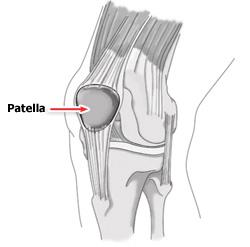
The patella, or kneecap lies in front of the knee joint. It is attached to the quadriceps muscle via the quadriceps tendon and acts to increase the leverage from this muscle group when straightening the knee. The patella glides up and down virtically within a groove called the patello-femoral groove at the front of the thigh bone(femur) during knee bending. Patella dislocation occurs when the patella moves outside of this groove. The patella may also sublux rather than fully dislocate, meaning it only moves partially out of position. These injuries  usually occurs as a result of an acute blow to or as a result of a twisting action on the knee. In most cases the patella will relocate into the patello-femoral groove on straightening of the knee. This, however is usually extremely painful. Risk factors which make a patella dislocation more likely are 1. insufficient quadriceps strength especially on the inside of the knee,   2. over pronation of the feet , and 3. an increased Q angle of the knee.

The vastus medialis obliqus muscle (VMO) is part of the quadriceps muscle group on the inside of the thigh, and is responsible for maintaining the stability of the patella towards the inside of the knee. If the VMO  isn’t strong enough, the patella is much more susceptible to dislocation.

Over pronation of the feet is where the feet roll in(collapse in slightly on the flattened arch  with the toes turning outwards) when running. This causes torsion at the knee, leaving the patella susceptible to moving outwards as the quadriceps contract.

The Q-angle of the knee is the angle formed by lines representing the pull of the quadriceps muscle and the axis of the patellar tendon. People with an increased Q ankle are often termed knock kneed. This angle when too large increases the risk of patellar dislocation.


 Treatment of patella dislocation

Apply  RICE(rest, ice, compression and elevation) principles to the injured knee . Ice can be applied for 10 to 15 minutes every hour initially reducing to 2 or 3 times a days as swelling and symptoms reduce.  Also medications like non steroidal anti-inflammatories (NSAIDs), analgesics, pain killers etc. would be beneficial, in order to manage both the inflammation and the pain .

Rest from any sporting activities and seek medical advice. An athlete who has had one episode of patella dislocation is often susceptible to another. The knee may need to be immobilized for 2-3 weeks initially. A knee support can provide protection for the joint initially during rehab. Patella stabilizing braces have been specifically designed to provide support for the knee cap.

Quadriceps strengthening is initiated during this phase. These should be in the form of static exercises. Quadriceps electrical stimulation is an option for muscle reeducation if the patient has difficulty activating the muscle secondary to pain. Electrical stimulation may also play a role in the management of knee joint effusion. When swelling, inflammation has subsided, the patient may progress to the recovery phase of rehabilitation.

When the acute symptoms settle down a full knee strengthening rehabilitation program is required to help avoid future recurrence. Specifically exercises for the VMO muscle on the inside of the quadriceps are important, but overall quadriceps strengthening is also beneficial . The VMO is an important medial stabilizer of the patella.  The prevailing theory has been that lateral patellar tracking is associated with VMO weakness. However, research has been inconclusive for VMO weakness as a direct causative mechanism of lateral patellar tracking(patellar subluxation/dislocation). Recent findings have shown that general quadriceps strengthening has demonstrated reductions in lateral tracking irrespective of the mechanism. Therapy should also include a protocol for hamstring muscle stretching. Tight hamstring muscles functionally counteract their agonist group, the quadriceps.


Any physical therapy program for patellofemoral problems must address any tightness or reduced flexibility in the quadriceps, hamstrings, hip abductors,  iliotibial band and calf muscles.

Medial patellar gliding exercises may loosen lateral retinacular tightness in this stage. Medial patellar gliding exercises are performed with the leg extended. The patient manually pushes the patella medially and holds for a count of 10 seconds.

An important concept in the rehabilitation of patellar dislocation and patellofemoral pain is knee flexion. Initially, any activity that requires greater than 40-45° of knee flexion causes symptoms. Initial rehabilitation programs start with the isometric open kinetic chain . Early rehabilitation programs should limit all activities that require quadriceps firing with the knee flexed greater than 45°.

Once isometric open kinetic chain exercises are tolerated without discomfort, the rehabilitation program advances to closed kinetic chain exercises (eg, mini squats, lunges, stair climbing). The rectus femoris, VMO, and VL are all strengthened by the mini squats (repetitions and sets modified to the tolerance of the patient).

Important goals are to restore ”range of movement” in the joint, mobilize soft tissues, and strengthen the surrounding musculature. Lunges and bike riding allow strengthening through a controlled ”range of movement”.  Once pain has resolved sufficiently to complete daily activity requirements without exacerbations, the patient can advance to the final phase of rehabilitation.

Advanced rehabilitation programs progress to jogging, running, plyometrics, and sport-specific exercises. Patients must be monitored and must always follow proper technique, as well as learn to properly fire the VMO.

Sometimes Surgery is needed. Most surgeons agree, if the kneecap dislocates multiple times, then surgery should be considered. When the kneecap dislocates, it is possible to damage the cartilage in the knee, leading to an increased risk of knee arthritis. When multiple dislocations occur, patellar stabilization surgery should be considered. While dislocations of the patella can occur after surgery, they are much less common.

Physiotherapist in Tralee

bicipital-tendonitis
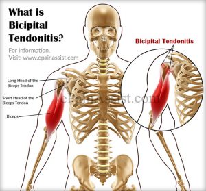
Bicipital tendonitis is a common cause of shoulder pain, often developing in people who perform repetitive, overhead movements. Biceps tendinitis develops over time, the pain being located at the front of the shoulder. The biceps muscle has two parts referred to as the long head and the short head. The tendon of the long head of the biceps is most commonly implicated with tendonitis. When this tendon is subjected to repetitive stresses, it can become irritated, swollen, and painful.This occurs where the tendon sits within the bicipital groove at the top of the humerus under the transverse ligament before it becomes part of the shoulder joint capsule.

Pain at this exact spot when pressed with a finger as the arm is rotated in and out while standing, is usually a fairly reliable test to confirm this condition. Imaging techniques such as MRI are typically not needed to diagnose biceps tendonitis.

Symptoms – Bicipital Tendonitis

Pain or tenderness in the front of the shoulder, which worsens with overhead lifting or activity.

Pain that moves down along the upper arm

An occasional snapping sound or sensation in the shoulder

Treatment – Bicipital Tendonitis

The initial goals of treatment for bicipital tendonitis are to reduce inflammation and swelling. Patients should restrict above shoulder height movements, reaching out with the affected arm and lifting. They should apply ice to the affected area for 10-15 minutes, 2-3 times daily for several days. Non-steroidal anti-inflammatory drugs (NSAIDs), such as ibuprofen, may help recovery. Probably the biggest aid to recovery is rest from the aggravating activity for several weeks. I often come across this condition in weight trainers who front press or incline bench press, the bar being too far out from their neck during pressing. It is also common in swimmers with poor technique or who ramp up their training distance/pace too quickly.


Physiotherapist Tralee : Phone 086-7700191 for an appointment, second opinion or to discuss your injury.

Lumbosacral Plexopathy

Lumbosacral Plexopathy : Recently I had a patient present with footdrop (foot drops and drags as you walk due to weakness in dorsiflexor muscles), also severe pain down the leg, along with lumbar-sacral pain(lower back area). Initially this might appear to be signs of disc herniation with nerve root compression, even though the footdrop would be fairly uncommon with that condition. An MRI showed the spine to be normal. We also looked at anything that might have injured the peroneal nerve which is quiet close to the skin on the lower leg, which in itself could cause the footdrop, but there was no sign of injury. We were also able to rule out diabetes which can lead to nerve damage.

So where did we go next? Answer : An electromyography (EMG)(nerve conduction study). Electromyography is a diagnostic procedure used to assess the health of muscles and the nerve cells that control them (motor neurons). EMG results can reveal nerve dysfunction, muscle dysfunction or problems with nerve-to-muscle signal transmission. As a result of this testing by a specialist, the client was diagnosed as having a Lumbosacral plexopathy. In this case it seemed that the nerve had been damaged by a viral or bacterial infection. More tests need to be done to confirm and treat this infection.

The lumbosacral plexus represents the nerve supply to the lower back, pelvis and legs. Lumbosacral plexopathy is characterized as inflammation and or damage to the nerve bundles in the area of the lumbar and sacral vertebrae regions. Symptoms may include shooting or burning pain, numbness, and decreased movement in the thigh, buttock and/or leg area. It is an uncommon idiopathic disorder(relates to or denotes any disease or condition which arises spontaneously or for which the cause is unknown). It is characterized by the acute onset of severe lower extremity pain followed by wasting and weakness of leg muscles along with variable sensory loss.

To Summarise : Lower Lumbosacral Plexopathy

It predominantly affect the L4–S3 nerve fibers.

There patient complains of a deep boring pain in the pelvis that can radiate posteriorly into the thigh and down into the posterior and lateral calf.

The ankle reflex may be depressed or absent.

Sensory symptoms and signs may be severely deminished over the posterior thigh and posterior-lateral calf and in the foot.

Other Causes of Lumbosacral Plexopathy

Structural
Retroperitoneal hemorrhage (anticoagulation, hemophilia)
Pelvic or abdominal tumor
Aneurysm (common or internal iliac artery)
Endometriosis
Trauma
Nonstructural
Inflammatory (plexitis)
Infarction
Postpartum [during childbirth]
Diabetes (diabetic amyotrophy – most common non-structural cause)
Radiation
Postsurgical (retractor injury)

Please note that the presentation of Lumbosacral Plexopathy has subtle differences to sciatica and discogenic pain, the later conditions being much more common than Lumbosacral Plexopathy.


Physiotherapists in Tralee  Phone 086-7700191 to discuss your condition or to make an appointment.

Phones, laptops, reading in bed and the consequences of poor posture.

Poor PostureThe increasing time we are spending glued to our phones or in front of a laptop with poor posture is manifesting itself with a large increase in the incidence of neck and shoulder pain presenting at clinic. Even kids are suffering from it. Good posture, as it relates to the neck, and outlined by a physiotherapist is commonly considered to be when the ears are positioned directly above the shoulders with the chest open and shoulders back. This neutral position minimizes the stress on the neck(cervical spine). Also it is important that the head is turned to full rotation left and right regularly in order to keep joints and muscles mobile and supple.

Imbalances from poor posture

Forward head posture occurs when the neck slants forward, placing the head further in front of the shoulders rather than directly above. This can lead to problems such as


  • Increased stress on the cervical spine. As the head moves forward in poor posture, the cervical spine must support an increased amount of weight.
  • Muscle overload. Forward head posture means muscles of the neck, shoulders and upper back have to continually overwork to counterbalance the pull of gravity of the forward head. As a result, muscles become more susceptible to painful strains and spasms.
  • Hunched upper back. Forward head posture is often accompanied by forward shoulders and a rounded upper back, which can increase the risk of developing pain and dysfunction in the neck, upper back, and/or shoulders.

The above can lead to more severe manifestations if the forward head posture is maintained long term. These include

  • Muscle imbalances. Some muscles in the neck, upper back, shoulders, and chest can become shortened and tight, whereas others can become elongated and weak.
  • Elevated risk for spinal degeneration. Extra stress on; the discs of the cervical spine’s, the facet joints, and vertebrae increases the risk of degenerative spinal issues, such as cervical degenerative disc disease and cervical osteoarthritis. It can also contribute to the development of bone spurs.
  • There is an increased risk of development of shoulder bursitis, specifically subacromial bursitis
  • Reduced mobility. With increased stiffness in the muscles and/or joints, the neck’s range of motion becomes decreased.
  • Nerve pain. Spinal degeneration and muscle spasm in the neck can lead to nerve compression resulting in neurogenic pain. This pain can be severely debilitating.

Treatment

Physiotherapist treatment of the above may consist of deep tissue massage, mobilizations and posture correction exercises. Severe cases of disc degeneration and/or nerve compression may require surgery. Mostly people suffer from the effects of poor posture later in life. The effects take time to manifest. Often people will only get the message of how detrimental poor posture can be when they start suffering from neuropraxia (nerve compression/ pain,tingling, numbness running down yours arms, worse at night). This condition can be quiet challenging for a physiotherapist treat. Physiotherapy treatment can still be very successful if it is caught in the early stages.


Physiotherapist in Tralee.

Back Pain Treatment Options Made Simple

Three very common causes of back pain presenting in clinic are 1. muscular, 2. discogenic (from a disc), 3. locked facet joint. We will look at each and the relevant back pain treatment options.

Muscular back pain

For the low back a common muscle to cause trouble is the piriformis muscle (see diagram). It is involved in hip rotation movements so if it tightens you lose rotation putting pressure on the lower back during activities. When the piriformis muscle tightens it may also squeeze on the sciatic nerve which travels from the lumbar spine  down the back of the leg causing pain anywhere along the nerve path(low back, hamstring, calf, sole of foot). Tightened hamstrings may also contribute to low back pain(often resulting from being seated for long periods) making the spine do more work due to resulting poor posture in lifting etc.

The piriformis muscle can usually be worked out and loosened  with deep tissue massage.  The hamstrings can also be worked if these are contributing. Electro-acupuncture also helps loosen the muscles/trigger points and settle nerve irritation in affected areas.

Back pain

Locked Facet joint back pain

This is a common cause of back pain in the upper thoracic region(along back of the rib cage)…Often the person feels like somebody is sticking a finger into their back when they inhale deeply. Also with this condition a person may find that they can turn completely in one direction but not the other. In more serious cases pain may shoot down the arm due to nerve entrapment/irritation. A locked facet joint may also occur in the lower back sending nerve pain down the leg. The crack you hear when the back is manipulated is locked facet joints opening, it is not a ”slipped disc” being put back into place. A timely spinal manipulation done correctly, with a little soft tissue work done thereafter can often sort this condition. With the manipulation you get a little crack if the facet joint was locked, as it opens, giving immediate relief.

Back pain


Discogenic (Disc) related back pain

This is a more tricky one. Probably most common in the lower back (lumbar region). The patient will sometimes present with their spine all twisted off to one side in a sort of ”S” shape. This is usually because the disc bulge or protrusion is touching a nerve. The body compensates for this by trying to get the disc away from the nerve, hence the ”S” shape. Also there will be muscle spasm as the body tries to protect itself. A patient with this condition is usually in a lot of pain and even simple tasks like walking can be excruciatingly painful. Disc pain is less common in the thoracic region but again here it presents with deep unrelenting pain.

There is no such thing as a ”slipped disc”, it is just a misnomer. Discs basically bulge, degenerate(collapse) or herniate(a little fluid filled sack can protrude or leak out of them)(see diagram below). It is when this bulge or protrusion touches a nerve, the trouble starts.

Basically a muscle relaxant (valium) from your doctor along with an anti-inflammatory (NSAIDS) for a couple of days usually helps a lot.  Usually two treatment sessions along with the medication to loosen out the muscles will also reduce pain and improve recovery time. A manipulation to open any locked facet joints may help as the condition settles.

When a disc touches a nerve it initiates a series of reactions including muscle spasm and inflammation. The medication is very important in this condition and the combination of the both muscle relaxant and anti-inflammatory seem to complement each other along with treatment. Things such as ”Mckenzie technique” and ”nerve glides” can also help when introduced at the right time during treatment but require too much detail to go into here.

Once the condition settles it is important to 1. Train your body to engage its core. 2. Strengthen your core muscles with pilates type exercises, along with developing your core fitness. 3. Lose weight if you are overweight. 4. Try and improve your flexibity.

Once you get somebody moving, loosened out, give them a proper rehab routine and they are taking the medication, it is more about giving the body time to heal itself than anything else. Basically what you are trying to do with initial treatment is to relax any muscle spasm, reduce inflammation, reduce pressure on the disc with the more long-term goal of strengthening the body in the hope of bringing back in the disc bulge/ protusion enough from the nerve it is touching, so that symptoms subside. If there is little improvement in your condition after a week, an mri may be required to get more accurate information on the injury.  In some cases an epidural or surgery  may be required.

Back pain

Please note the above article only lists some common causes of back pain.

Back pain treatment in Kerry –  phone 086-7700191