equinus

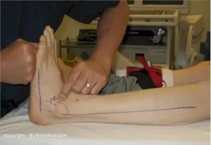
Equinus Foot Condition

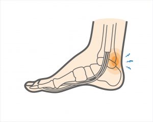
The ankle joint connects the leg to the foot. It is formed by three separate bones, the tibia, fibula and talus. The shinbone (tibia) supports most of a person’s weight when standing. The outer bone (fibula) is the smaller bone of the lower leg. A small, irregular-shaped foot bone (talus) connects the tibia and fibula. Acting as a hinge, these bones form the ankle. The ankle joint allows movement such as walking, running and jumping, and also contributes to lower limb stability.

The ankle is reinforced by ligaments which connect bone to bone. Ligaments have a mildly elastic structure that allows them to stretch, within their limits, and then return to their normal positions. Ligaments protect the ankle from abnormal movements—especially twisting, turning and rolling of the foot.

Description

A person with equinus has a limited range of ankle motion and lacks the flexibility needed to bring the top of the foot upward, toward the shin. It may be either congenital or acquired, and occurs equally in both men and women.  Equinus can be due to several different reasons including the following:

• bony block between the talus and distal tibia (osseous equinus);
• contracture or tightness of the soleus muscle (inner calf muscle);
• contracture or tightness of the soleus and gastrocnemius muscles (gastroc-soleal equinus);
• isolated tightness of the gastrocnemius muscles (outer calf muscle); and
• compensatory loss of ankle joint range of motion for some other condition such as pes cavus (pseudoequinus).


People with equinus often develop ways to compensate for their limited ankle motion. Depending on how a patient compensates for the inability to bend properly at the ankle, a variety of other foot conditions can develop, such as:

  • Plantar Fasciitis
  • Calf cramping
  • Achilles Tendinitis
  • Metatarsalgia (pain and/or callusing on the ball of the foot)
  • Flatfoot
  • Arthritis of the midfoot (middle area of the foot)
  • Pressure sores on the ball of the foot or the arch
  • Bunions and hammertoes
  • Ankle pain
  • Shin splints
  • Sesamoiditis
  • Hallux valgus
  • hallux rigidus
  • Hammer toes


Nonsurgical Treatment of Equinus

Some nonsurgical treatment strategies are aimed at relieving the symptoms and conditions associated with equinus. Treatment for the equinus itself may include one or more of the following options.

  • Heel lifts—Placing heel lifts inside the shoes or wearing shoes with a moderate heel may reduce symptoms by taking stress off the Achilles tendon and compensating for the restricted movement of the ankle joint. The joint is meant to have about fourteen degrees of movement ideally, so say it has only four degrees of movement, adding a ten degree heel lift helps compensate for the missing degrees of movement .
  • Arch supports or orthotic devices—Custom orthotic devices that fit into the shoe are often prescribed to ensure that weight is distributed properly, and to help control muscle/tendon imbalance. Again these devices will most likely include a heel lift.
  • Physical therapy—To help remedy muscle tightness, deep tissue massage of calf muscles along with a stretching program for the calf muscles are recommended.


For a quick simple video describing the condition click   here


See our wide range of orthotics  here


Posterior ankle impingement physiotherapyPosterior ankle impingement is a condition characterised by tissue damage at the back of
the ankle joint due to compression of these structures. This occurs when the foot and ankle
are pointed maximally away from the body (plantarflexion – figure 1. ). It may occur when
compressive forces are too repetitive and/or too forceful. This can occurs in the presence of
ankle swelling or bony anomalies, such as additional bone, a condition known as an “os
trigonum”. Posterior ankle impingement is most commonly found in gymnasts, ballet
dancers, and footballers, because they regularly maximally plantarflex their ankles during
their activities. The condition can also occur due to inadequate rehabilitation of an acute
ankle injury (ie. ankle sprain).

Mechanism of Injury

Posterior ankle impingement may develop due to an acute traumatic plantar hyperflexion
event, such as an ankle sprain. It may also occur as a result of repetitive low-grade trauma
associated with plantar hyperflexion, say like in case of a female ballet dancer. It is
important to differentiate between these two, because the latter, that is posterior
impingement from overuse, has a better prognosis.
The anatomy of the posterior ankle is a key factor in the occurrence of posterior
impingement syndrome . The more common causes of the condition are osseous in nature,
such as the os trigonum, an elongated posterolateral tubercle of the talus (known as
Stieda’s process), a downward sloping posterior lip of the tibia, an osteophyte from the
posterior distal tibia , or a prominent posterior process of the calcaneus. However, posterior
impingement can also be soft tissue related, as with a thickened posterior joint capsule ,
post-traumatic scar tissue, post-traumatic calcifications of the posterior joint capsule, or
loose bodies in the posterior part of the ankle joint. Symptoms for all of these conditions
relate to physical impingement of osseous or soft tissue structures, resulting in painful
limitation of the full range of ankle movement.
The most common cause ''os trigonum'' is an extra (accessory) bone that sometimes
develops behind the ankle bone (talus). The mineralized os trigonum appears between the
ages of 7 and 13 years and usually fuses with the talus within 1 year, forming the trigonal
(Stieda) process. It may remain as a separate ossicle in 7-14% of patients, and is often
bilateral(in both ankles). An os trigonum can be a focus of osseous abutment against other
structures. Pain can also be caused by disruption of the cartilaginous synchondrosis
between the os trigonum and the lateral talar tubercle as a result of repetitive microtrauma
and chronic inflammation.
In the case of soft tissue impingement it usually results from scarring and fibrosis associated
with synovial, capsular, or ligamentous injury ie. bad ankle sprain. It is thought that this
type of manifestation usually usually occurs when a significant soft-tissue component
forms. The soft-tissue component can consist of synovial thickening throughout the
posterior capsule or be more focal, involving the posterior intermalleolar or talofibular ligament. The flexor hallucis longus tendon runs in the groove between the lateral and
medial processes of the talus and can also be injured in posterior impingement, resulting in
tenosynovitis.


Signs and symptoms

Patients who have posterior impingement complain of chronic deep posterior ankle pain
worsened by forced plantar flexion or push-off forces as occur during activities such as
ballet dancing, jumping, or running downhill. In some patients, forced dorsiflexion(opposite
to plantarflexion) is also painful. Physical examination reveals pain on palpation over the posterolateral talar process, which is located along the posterolateral aspect of the ankle between the Achilles and peroneal
tendons . Passive forced plantar flexion results in pain and often a grinding
sensation as the posterolateral talar process is entrapped between the posterior tibia and
calcaneus.


Diagnosis of posterior ankle impingement

A thorough examination by an experienced practitioner may be all that is necessary to
diagnose posterior ankle impingement. Further investigations such as an X-ray, MRI, CT scan
or Ultrasound may help confirm diagnosis.


Physiotherapist in Tralee, Co. Kerry………..Phone 0867700191 to make an appointment or discuss your condition.

Suboccipital-Muscles-of-the-Neck

Suboccipital Muscles & Trigger Point Pain

Suboccipital muscles (see image) are a group of four muscles located on each side of the upper cervical spines, just below the base of the skull. The muscles connect the base of the skull with the top two vertebrae (C1 and C2) of the neck.

Poor posture in general especially with the increased use of portable electronic devices, which include mobile phones, laptops, and tablets has increased the prevalence of neck pain in both children and adults. Increased screen time on these devices is not only correlated with depression, sleep interruption, and poor food choices, but also rising rates of neck pain, especially in adolescents and young adults. This form of neck pain, including dysfunction of C1 and altered mechanics of the cervical spine due to poor posture, can also lead to headaches. Reading in bed is also a big offender. Any position where your head and neck are positioned forward and in a stationary position for long periods on time increases the likelihood of postural related spinal issues(ie. kyphosis, discogenic disorders etc.), neck pain and headaches.

These suboccipital muscles play an important role in controlling movements of your head and neck, providing sensory input and are also linked closely to vestibular and balance functions. However, when the suboccipital muscles become tightened, the following symptoms may occur. These could include:

  • Stiff neck
  • Neck pain
  • Headaches with a band of pain on the side of the head that extends from the back of the head to the eye as a result of active trigger points. This type of pain feels deep in the head, and often it is difficult to describe.

Messages sent to the brain may be altered, which is also why sometimes headache sufferers may also experience sensory symptoms, including dizziness and visual disturbances.


Physiotherapy Treatment

Treatment includes deep tissue work, trigger point release, manipulation, mobilisation, postural education and a rehabilitation program.


suboccipttal trigger points
Referred pain from suboccipital trigger points


Physiotherapists in Tralee Phone 0867700191


Overview of Shoulder Impingement by Physiotherapist in Tralee

shoulder-Impingement
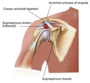
Shoulder impingement is a very common cause of shoulder pain, where a supraspinatus tendon inside the shoulder rubs or catches on nearby tissue and/or bone(acromium) as you lift your arm. Your shoulder is made up of several joints, muscles and tendons which allow the great range of motion in your arm. There are three bones in the shoulder joint: your upper arm bone, your shoulder blade, and your collarbone . Your arm is kept in your shoulder socket by your rotator cuff muscles, along with ligaments and the joint capsule which envelopes the shoulder. There is a lubricating sac called a bursa between the supraspinatus muscle of the rotator cuff where the supraspinatus travels under the bone on top of your shoulder (acromion). The bursa allows the supraspinatus tendon to glide freely under the acromium when you move your arm. When the bursa becomes swollen and inflamed, the condition is called bursitis and is one cause of shoulder impingement. Bone spurs within the shoulder joint can also contribute to impingement, as can forward curving shoulders due to years of bad posture. Sometimes due to impingement and/or bone spurs the supraspinatus muscle or tendon can tear. It can also happen during an accident like a shoulder dislocation or falling on a shoulder etc. Those who do repetitive lifting or overhead activities using the arm, such as paper hanging, construction, or painting are also susceptible.


Symptoms of shoulder impingement

You may have pain and stiffness when you lift your arm. There may also be pain when the arm is lowered from an elevated position. Early symptoms may be mild. Patients frequently do not seek treatment at an early stage. These symptoms may include:

  • Minor pain that is present both with activity and at rest
  • Pain radiating from the front of the shoulder to the side of the arm
  • Sudden pain with lifting and reaching movements
  • Athletes in overhead sports may have pain when throwing or serving a tennis ball

As the problem progresses, the symptoms increase:

  • Pain at night
  • Loss of strength and motion
  • Difficulty doing activities that place the arm behind the back, such as buttoning or zippering

If the pain comes on suddenly, the shoulder may be severely tender. All movement may be limited and painful.


Imaging Tests

Other tests which may help your doctor confirm your diagnosis include:

X-rays. Becauses x-rays do not show the soft tissues of your shoulder like the rotator cuff, plain x-rays of a shoulder with rotator cuff pain are usually normal or may show a small bone spur. A special x-ray view, called an “outlet view,” sometimes will show a small bone spur on the front edge of the acromion.

Magnetic resonance imaging (MRI) and ultrasound. These studies can create better images of soft tissues like the rotator cuff tendons. They can show fluid or inflammation in the bursa and rotator cuff. In some cases, partial tearing of the rotator cuff will be seen.

Treatment

The goal of treatment is to reduce pain and restore function. In planning your treatment, your doctor will consider your age, activity level, and general health.

TreatmentIn cases where there is a tear in the supraspinatus muscle or tendon, conservative treatment such as rest, physio etc rarely works. In milder cases rest along with rehab and correction of postural problems in the shoulder may work.Rest. Your doctor may suggest rest and activity modification, such as avoiding overhead activities.

Non-steroidal anti-inflammatory medicines. Drugs like ibuprofen and naproxen reduce pain and swelling in mild cases.

Physical therapy. A physical therapist will initially focus on restoring normal motion to your shoulder. Stretching exercises to improve range of motion are very helpful. If you have difficulty reaching behind your back, you may have developed tightness of the posterior capsule of the shoulder (capsule refers to the inner lining of the shoulder and posterior refers to the back of the shoulder). Specific stretching of the posterior capsule can be very effective in relieving pain in the shoulder.

Once your pain is improving, your therapist can start you on a strengthening program for the rotator cuff muscles.

Steroid injection.  Cortisone is a very effective anti-inflammatory medicine. Injecting it into the bursa beneath the acromion can relieve pain and reduce symptoms dramatically if the sole cause of the impingement is bursitis.


Surgical Treatment

When nonsurgical treatment does not relieve pain, your doctor may reoved. This is also known as a subacromial decompression. These procedures can be performed using either an arthroscopic or open technique.

Arthroscopic technique. In arthroscopy, thin surgical instruments are inserted into two or three small puncture wounds around your shoulder. Your doctor examines your shoulder through a fiberoptic scope connected to a television camera. He or she guides the small instruments using a video monitor, and removes bone and soft tissue. In most cases, the front edge of the acromion is removed along with some of the bursal tissue.

Your surgeon may also treat other conditions present in the shoulder at the time of surgery. These can include arthritis between the clavicle (collarbone) and the acromion (acromioclavicular arthritis), inflammation of the biceps tendon (biceps tendonitis), or a partial rotator cuff tear.

Open surgical technique. In open surgery, your doctor will make a small incision in the front of your shoulder. This allows your doctor to see the acromion and rotator cuff directly.

Rehabilitation. After surgery, your arm may be placed in a sling for a short period of time. This allows for early healing. As soon as your comfort allows, your doctor will remove the sling to begin exercise and use of the arm.


Physiotherapists in Tralee, Co. Kerry for all your physiotherapy needs. Phone 0867700191

Temperomandibular Joint Disorder
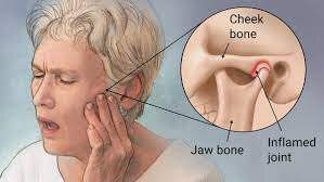
The temporomandibular joint works as a combination of hinge and sliding actions. It connects the jawbone to the skull on each side of the face.  The parts of the bones that interact in the joint are covered with cartilage and are separated by a small shock-absorbing disk, which normally keeps the movement smooth.  Temporomandibular  joint disorder (TMJD) can cause pain in your jaw joint and in the muscles that control jaw movement.
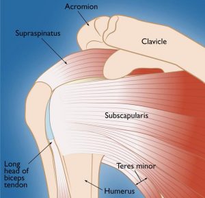
Symptoms

  • Pain in one or both of the temporomandibular joints
  • Difficulty or pain chewing
  • Locking of the joint, making it difficult to open or close your mouth
  • Clicking sound or grating sensation when you open your mouth or chew

Causes of  Temporomandibular Joint Disorder

Painful TMJ disorders can occur if:

  • The disk erodes or moves out of its proper alignment
  • The joint’s cartilage is damaged by arthritis
  • The joint is damaged by a blow or other impact

In many cases, however, the cause of TMJ disorders is unclear.

Treatment of  Temporomandibular Joint Disorder

In some cases, the symptoms of TMJ disorders may go away without treatment. Some of the following may also help.

Medications

  • Pain relievers and anti-inflammatories. These can help relieve pain and inflammation.
  • Muscle relaxants. These can help relax the jaw muscles.

Therapies

Nondrug therapies for TMJ disorders include:

  • Oral splints or mouth guards.  These devices worn at night while sleeping can help prevent grinding of the teeth.
  • Physical therapy.  Deep tissue massage work on neck and jaw muscles can be beneficial along with certain rehabilitation exercises.
  • Education. Education can help you understand the factors and behaviors that may aggravate your pain, so you can avoid them. Examples include teeth clenching or grinding, eating foods where you have to open the jaw wide like apples and burgers.

When conservative treatments fail, the following may be considered:

  • Arthrocentesis. This is a minimally invasive procedure that involves the insertion of small needles into the joint so that fluid can be irrigated through the joint to remove debris and inflammatory byproducts.
  • Injections. In some people, corticosteroid injections into the joint may be helpful. Infrequently, injecting botulinum toxin type A (Botox, others) into the jaw muscles used for chewing may relieve pain associated with TMJ disorders.
  • TMJ arthroscopy.  A small thin tube is placed into the joint space, an arthroscope is then inserted and small surgical instruments are used for surgery. TMJ arthroscopy has fewer risks and complications than open-joint surgery does, but it has some limitations as well.
  • Modified condylotomy. Modified condylotomy addresses the TMJ indirectly, with surgery on the mandible, but not in the joint itself. It may be helpful for treatment of pain and if locking is experienced.
  • Open-joint surgery. If your jaw pain does not resolve with more-conservative treatments and it appears to be caused by a structural problem in the joint, your doctor or dentist may suggest open-joint surgery (arthrotomy) to repair or replace the joint. However, open-joint surgery involves more risks than other procedures do.


For some handy self treatment tips, check out this video https://youtu.be/7b73yE0U2t0

Tmj manipulation video


Check out our website and feel free to contact us to discuss your condition or to set up an appointment.


When Back Pain Is A Symptom Rather Than A Condition

Back pain may sometimes be a symptom rather than a condition as such. A practitioner needs to always keep this at the back of their mind when treating or assessing somebody. Sometimes things need more investigation. A good physio will spot this early and refer you on quickly to your G.P. for further examination. It is always better to play it safe if in doubt.  I always believe you should be seeing improvement in your condition from treatment to treatment. If there is no improvement after several treatments you need to be asking yourself a few questions i.e. Do I need to try a different physio or do I need to consult my doctor for a second opinion. Sometimes the proper treatment protocol for a condition is all that is needed for a swift recovery. The list below is not meant to be alarmist. It is more to emphasize the fact that continuous unrelenting back pain can be a symptom of something else going on within your body.


Below are examples of conditions/medications etc. that may have back pain as a side effect/symptom.


kidney stones – A kidney stone may not cause symptoms until it moves around within your kidney or passes into your ureter (the tube connecting the kidney and bladder). At that point, you may experience symptoms such as severe pain in your side and back below the ribs, or pain that radiates into the lower abdomen and groin.

Lupus is a long-term autoimmune disease in which the body’s immune system becomes hyperactive and attacks normal, healthy tissue. Symptoms include inflammation, swelling, and damage to the joints, skin, kidneys, blood, heart, and lungs. Lupus can cause neck and back pain, because muscles in these areas can become inflamed due to the lupus. Furthermore, the muscle pain syndrome  ”fibromyalgia”  can cause pain in these areas and is commonly associated with lupus.

Spinal arthritis/facet joint arthritis causes stiffness and back pain.

Cancers – A primary bone cancer tumor in the spine can cause back pain, as can a number of other cancers when they have metastasized(spread to other sites in the body), such as breast cancer, testicular cancer, colon cancer, and lung cancer. In fact, back pain is often the one of first symptoms that people with lung cancer notice before they are diagnosed. A tumor in the lungs can put pressure on the spine, or can affect the nerves around the chest wall and spine.

Spondylosis a painful condition of the spine resulting from the degeneration of the intervertebral discs.

Spondylitis is a condition resulting in inflammation within the joints of the spine. As the inflammation goes and healing takes place, bone grows out from both sides of the vertebrae and may join the two together, causing a stiffening known as ankylosis. The progressed condition is called ankylosing spondylitis. The cause is not yet known.

back pain


Spondylolisthesis is a slipping of vertebra that occurs, in most cases, at the base of the spine.

Spondylolysis is a defect or fracture of one or both wing-shaped parts of a vertebra, can result in vertebrae slipping backward, forward, or over a bone below.


Spondylosis, Spondylitis, Spondylolisthesis, Spondylolysis all have back pain as a symptom.


Fractures – Even a minor fracture along the spine  can cause considerable back pain.

StatinsStatins are drugs that can help lower your cholesterol. One of the more severe side effect of statins is myotoxicity(having a toxic effect on muscle), in the form of myopathy(a disease of the muscle in which the muscle fibers do not function properly. This results in muscular weakness), myalgia(muscle pain), myositis(inflammation and degeneration of muscle tissue) or rhabdomyolysis(a condition in which damaged skeletal muscle breaks down). Currently, the only effective treatment of statin-induced myopathy is the discontinuation of statin use in patients affected by muscle aches, pains and elevated creatine kinase levels. Creatine kinase are the clinical measure of muscle damage (rhabdomyolysis).

Anticonvulsant drugs such those used in the treatment of conditions like epilepsy can cause changes in calcium and bone metabolism. This may in time lead to decreased bone mass and a risk of osteoporotic fractures in the spine which may also result in severe back pain. Two widely used antiepileptic drugs phenytoin and carbamazepine are recognized to have direct effects on bone cells.

Corticosteroids – Longterm use of corticosteroids increase the risk of compression fractures in the spine(back).


Neurological Compromise — A Red Flag

Neural compromise can result from spinal cord or cauda equina compression . Cauda equina compression usually results from a fracture, tumor, epidural hematoma, or abscess, and occasionally from a massive disk herniation. Paraplegia(impairment in motor or sensory function of the lower extremities), quadriplegia(paralysis caused by illness or injury that results in the partial or total loss of use of all four limbs and torso), or cauda equina deficit should trigger an aggressive search for the cause.

Cauda equina compression classically presents with back pain, bilateral sciatica(pain down the back of both legs), saddle anesthesia, and lower extremity weakness progressing to paraplegia, but in practice these symptoms are variably present and diagnosing the condition often requires a high degree of suspicion. Hyporeflexia(no reflexes) is typically a sign of cauda equina compression, while hyperreflexia(overresponsive reflexes), clonus(series of involuntary, rhythmic, muscular contractions and relaxations), and the Babinski sign(see below *)  suggest spinal cord compression, requiring an evaluation of the cervical and thoracic spine. Cauda equina compression typically involves urinary retention; in contrast, cord compression typically causes incontinence. If either cauda equina or spinal cord compression is detected during an initial examination, an immediate more extensive evaluation is warranted. MRI is the study of choice.

*(The Babinski reflex occurs after the sole of the foot has been firmly stroked. The big toe then moves upward or toward the top surface of the foot. The other toes fan out. This reflex is normal in children up to 2 years old. It disappears as the child gets older. It may disappear as early as 12 months. When the Babinski reflex is present in a child older than 2 years or in an adult, it is often a sign of a central nervous system disorder. The central nervous system includes the brain and spinal cord.)

Spinal epidural hematoma

Spinal epidural hematoma is a rare but dramatic cause of paralysis in elderly patients. In most cases, there is no antecedent trauma. Lawton et al. (1995), in a series of 30 patients treated surgically for spinal epidural hematoma, found that 73% resulted from spine surgery, epidural catheterization, or anticoagulation therapy. Other possible causes of epidural hematoma include vascular malformations, angiomas, aneurysms, hypertension, and aspirin therapy. The same study found that the time from the first symptom to maximal neurologic deficit ranged from a few minutes to 4 days, with the average interval being nearly 13 hours.

Although painless onset has been reported, spinal epidural hematoma typically presents with acute pain at the level of the lesion, which is often rapidly followed by paraplegia(impairment in motor or sensory function of the lower extremities) or quadriplegia(is paralysis caused by illness or injury that results in the partial or total loss of use of all four limbs and torso), depending on the location of the hemorrhage. Sometimes the onset of pain is preceded by a sudden increase of venous pressure from coughing, sneezing, or straining at stool. Urinary retention often develops at an early stage.

Most lesions occur in the thoracic region(rib area of back) and extend into the cervicothoracic(upper back and neck) or the thoracolumbar(ribs to lower back area) area. The pain distribution may be radicular(affecting or relating to the root of a spinal nerve), mimicking a ruptured intervertebral disk.

Evaluation should be with MRI. Early recognition, MRI confirmation, and treatment should be accomplished as soon as possible. Recovery depends on the severity of the neurologic deficit and the duration of symptoms before treatment. Lawton et al.(1995), found that patients taken to surgery within 12 hours had better neurologic outcomes than patients with identical preoperative neurologic status whose surgery was delayed beyond 12 hours. Surgery should not be withheld because of advanced age or poor health: in 10 reported cases in which surgery was delayed, all patients died.


For more about treatments and opening times etc. click on our homepage

ROTATOR CUFFRotator Cuff Tears

Your arm is kept in your shoulder socket by your rotator cuff. The rotator cuff consists of four muscles, the supraspinatus muscle, the infraspinatus muscle, the terres minor muscle, and the subscapularis muscle. These muscles attach the humerus to the shoulder blade and help lift and rotate your arm.

Your shoulder is made up of three bones: your upper arm bone (humerus), your shoulder blade (scapula), and your collarbone (clavicle). The shoulder is a ball-and-socket joint: the ball, or head, of your upper arm bone fits into a shallow socket in your shoulder blade.

There is also a lubricating sac called a bursa between the rotator cuff muscles and the bone on top of your shoulder (acromion). The bursa allows the rotator cuff tendons to glide freely when you move your arm. Sometimes it may become inflamed and painful due to overuse.


Rotator Cuff Tears – Causes

There are two main causes of rotator cuff  injury and degeneration.


1. Acute Tears

If you fall down on your outstretched arm or lift something too heavy with a jerking motion, you can tear your rotator cuff at that moment.

2. Degenerative Tears

Many rotator cuff tears are the result of a wearing down of the tendon occurring slowly over time. They are more common in the dominant arm.

Several factors contribute to degenerative, or chronic, rotator cuff tears.

  • Repetitive stress : Repeating the same shoulder motions again and again can stress your rotator cuff muscles and tendons. Baseball, tennis, rowing, and weightlifting are examples of sports activities that can put you at risk for overuse tears. Many jobs and routine chores can cause overuse tears, as well.
  • Lack of blood supply : As we get older, the blood supply to our rotator cuff tendons reduces. Without a good blood supply, the body’s natural ability to repair tendon damage is impaired. This can ultimately lead to a tendon tear.
  • Bone spurs : As we age, bone spurs (bone overgrowths) often develop on the underside of the acromion bone. When we lift our arms, the spurs rub on the rotator cuff tendon. This is part of a condition which comes under the general term of ”shoulder impingement”.  Over time this can damage the tendon, making a tear more likely. The supraspinatus muscle is probably the more common of the rotator cuff muscles to get injured as we age. This is because the supraspinatus passes under the acromion process before attaching to the greater tubercle of the humerus.
  • Poor posture : With poor posture over time the shoulders start to protrude forward making the subacromion space smaller and causing shoulder impingement, sometimes leading to rotator cuff muscle/tendon damage, especially the supraspinatus muscle/tendon.

 Symptoms

The most common symptoms include:

  • Pain at rest and at night, particularly if lying on the affected shoulder
  • Pain when lifting and lowering your arm or with specific movements
  • Weakness when lifting or rotating your arm

Tears that happen suddenly, such as from a fall, usually cause intense pain immediately. There may be a snapping sensation or feeling of something giving way, followed by immediate weakness/severe pain in moving your upper arm.Tears that develop slowly due to overuse also cause pain and arm weakness but the increase in pain intensity occurs over time. You may have pain in the shoulder when you lift your arm, or pain that moves down your arm. At first, the pain may be mild and only present when lifting your arm over your head, such as reaching into a cupboard. Over time, the pain may become more noticeable at rest, and no longer goes away with medications. You may have pain when you lie on the injured side at night. The pain and weakness in the shoulder may make routine activities such as combing your hair or reaching behind your back more difficult.

There are different types of tears.

  • Partial tear : This type of tear is also called an incomplete tear. It damages the tendon, but does not completely sever it.
  • Full-thickness tear : This type of tear is also called a complete tear. It separates all of the tendon from the bone.


Rotator Cuff Tears – Imaging Tests

Magnetic resonance imaging (MRI) or ultrasound are probably the best way to confirm tears, bursitis, tendinopathies or bone spurs in the rotator cuff area.

 Rotator Cuff Tears -Treatment

If you have a partial rotator cuff tear and you keep using it despite increasing pain, you may cause further damage.  A rotator cuff tear can get larger over time.

Chronic shoulder and arm pain are good reasons to see your doctor. Early treatment can prevent your symptoms from getting worse. It will also get you back to your normal routine that much quicker.

The goal of any treatment is to reduce pain and restore function. There are several treatment options, and the best option is different for every person. In planning your treatment, your doctor will consider your age, activity level, general health, and the type of tear you have.

Nonsurgical Treatment

Nonsurgical treatment options may include:

  • Rest. Your doctor may suggest rest and limiting overhead activities. He or she may also prescribe a sling to help protect your shoulder and keep it still.
  • Activity modification. Avoid activities that cause shoulder pain.
  • Nonsteroidal anti-inflammatory medication. Drugs like ibuprofen and naproxen reduce pain and swelling.
  • Strengthening exercises and physical therapy. Specific exercises will restore movement and strengthen your shoulder. Your exercise program will include stretches to improve flexibility and range of motion. Strengthening the muscles that support your shoulder can relieve pain and prevent further injury.
  • Cortisone steroid injection. This is not recommended with a muscle or tendon tear and can actually lead to more damage in the long-term. One exception would be in the case of a subacromial bursitis where it can be very effective in shrinking the bursa back to normal size and reducing the inflammation.

Surgical Treatment

Your doctor may recommend surgery if your pain does not improve with nonsurgical methods. Continued pain is the main indication for surgery. If you are very active and use your arms for overhead work or sports, your doctor may also suggest surgery.

Other signs that surgery may be a good option for you include:

  • Your symptoms have lasted 6 to 12 months
  • You have a large tear
  • You have significant weakness and loss of function in your shoulder
  • Your tear was caused by a recent, acute injury

Surgery to repair a torn rotator cuff most often involves re-attaching the tendon to the head of humerus (upper arm bone).

Home rehab exercises for the rotator cuff muscles click here


Physiotherapist in Tralee : Phone 086-7700191

Gout ?

Gout
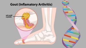
Gout is a form of arthritis which starts as a result of excess uric acid build-up in the blood, also called hyperuricemia. It is an extremely painful condition. Uric acid is produced in the body during the breakdown of purines – chemical compounds that are found in certain foods such as meat, poultry, and seafood. Normally, uric acid is dissolved in the blood and is excreted from the body in the urine via the kidneys. If too much uric acid is produced, it can build up and form needle-like crystals that trigger inflammation and pain in the joints and surrounding tissue. The condition often starts by affecting the joint at the base of the big toe. Attacks often occur without warning and in the middle of the night. They can come on quickly and keep returning over time, slowly damaging tissues in the region of the inflammation.

Tests and diagnosis

Gout can be tricky to diagnose, as it’s symptoms can be similar to those of other conditions, i.e. bunion on the big toe. While hyperuricemia occurs in the majority of people that develop gout, it may not be present during a flare. There is one very subtle, important difference between gout and other foot conditions, that helps the health practitioner differentiate between them. When you very gently stroke the skin of a gout affected area, it will cause a lot of pain in the sufferer. This does not occur with other conditions. The gout makes the skin super sensitive. This test is not 100% accurate, but I have found it to be a reasonably good indicator that one should look in the direction of gout, and refer the patient onto a doctor. This is especially true, if during examination and history take, the patient meets other criteria that would make them more prone to gout, and there has been no specific injury to the area affected. Often with tests you are more trying to rule out other conditions and narrow your possibilities down to that of gout..

One diagnostic test that doctors can carry out is the joint fluid test, where fluid is extracted from the affected joint with a needle. The fluid is then examined to see if any urate crystals are present. As joint infections can also cause similar symptoms to gout, a doctor can also look for bacteria when carrying out a joint fluid test in order to rule out a bacterial cause. Doctors can also do a blood test to measure the levels of uric acid in the blood. However, people with high uric acid levels do not always experience gout. Equally, some people can develop the symptoms without having increased levels of uric acid in the blood. Finally, doctors can search for urate crystals around joints or within a tophus(deposit of uric acid crystals) using ultrasound or CT scans.  X-rays cannot detect gout, but may be used to rule out other causes.

Treatment

Treatment and control of the condition requires certain dietary modifications such as reducing the intake of foods high in purines ie. red meat, shellfish, and beer to name but a few. Other factors that contribute to gout include being overweight, certain medications, high blood pressure, drinking too many sugary sodas, and being regularly dehydrated. These all need to be monitored and corrected.

Treatments for acute attacks include:

  1. NSAIDs – Non-Steroidal Anti-Inflammatory Drugs
  2. Colchicine
  3. Steroids
  4. Urate-Lowering Therapy (ULT)


For more see video


Overview by physiotherapists in Tralee, Co. Kerry


Lumbar Spinal Stenosis

spinal stenosis
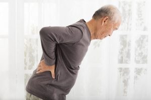
Spinal stenosis is a narrowing of the space within the spine. This can put pressure on the nerves that travel through the spine. It occurs most often in the lower back and the neck. Some people are born with a congenital form, but most develop it as part of the degenerative cascade. Sometimes people do not feel effects of the narrowing, but as part of the aging process, most people will eventually notice radiating pain, weakness, and/or numbness secondary to the compression of the nerves or spinal cord. In lumbar stenosis, the spinal nerve roots in the lower back become compressed and this can produce – tingling, weakness or numbness that radiates from the low back and into the buttocks and legs, especially with activity.

Lumbar spinal stenosis often mimics the symptoms of vascular insufficiency. Both conditions can cause claudication, which means leg pain with walking. If vascular studies identify normal blood flow, and tests confirm spinal stenosis,the symptoms are then referred to as neurogenic claudication.


myofascial-pain-syndrome
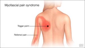
Myofascial Pain Syndrome

Myofascial pain syndrome is where pressure on sensitive points within your muscles (trigger points) causes pain and sometimes refers pain to seemingly unrelated parts of your body. It can occur after a muscle has been contracted repetitively ie. repetitive motions used in jobs or hobbies or by stress-related muscle tension. While nearly everyone has experienced muscle tension pain at some point, the discomfort associated with myofascial pain syndrome persists or worsens. There is no laboratory, radiographic or other diagnostic tests to prove the diagnosis of myofascial pain syndrome so it is considered a “subjective” diagnosis. Myofascial trigger points do not always cause pain. Sometimes they can lie dormant or inactive within a muscle for months or even years. Trigger points can often be identified by a skilled therapist working through your muscles with deep tissue massage. They feel like little knots deep within the muscles which when pressed are extremely tender.

Fascia is the body’s connective tissue. It is a head to toe, all-encompassing and interwoven system of fibrous connective tissue found throughout the body. Your  fascia provides a framework that helps support and protect individual muscle groups, organs, and the entire body as a unit. It is the same as that cling film, elastic type structure that surrounds a joint of meat, helping hold it together when the outer skin is removed.

This fascia in itself can also contribute to ”myofascial pain” syndrome. Injury, illness, stress, aging and repetitive use, can cause the fascia to shorten, thicken and become more unyielding . All the nerves and blood vessels run through the fascia. Therefore, if this connective tissue is tight, the associated tissues will have poor nutrient exchange. This exacerbates any painful situation because toxic metabolic waste products build up which often further aggravate pain receptors. This can create a vicious cycle, leading to increased muscle tension and further thickening and hardening of the fascia, which in turn further limits mobility.

 Symptoms

Signs and symptoms of myofascial pain syndrome may include:

  • Deep, aching pain in a muscle
  • Pain that persists or worsens
  • A tender knot in a muscle
  • Difficulty sleeping due to pain
  • Spasm in the area

The most commonly affected muscle groups include those of the neck, shoulders, upper  & lower back. Generally one side of the body is more affected than the other. It is common for patients with myofascial pain syndrome to have poor sleep patterns. This is associated with feeling unrested after a nights ”sleep” and daytime tiredness . Stiffness after inactivity is also a common feature.


Treatment

Myofascial pain can often benefit greatly from ”proper skilled deep tissue massage”, and trigger point release, both manual and with dry needling techniques. The fascia can also be stretched and worked out during the massage.  This is also one of the benefits of a stretching program when incorporated into your exercise routine. It helps keep prevent the fascia from tightening up because you are continually stretching it out.

During treatment for myofascial pain it is important that the patients reduce their stress levels, if this is a contributing factor to the condition. Exercises prescribed by a physio may also help, along with improving ones sleep patterns. In severe, chronic cases, medications may be needed to aid recovery.

Often trials of different medications are used to find the best treatment for a particular patient. For example, trazodone or amitriptyline may be used at bedtime to improve sleep as well as relieve pain;  cyclobenzaprine or  orphenadrine can also be  used to relax muscles and aid sleep; and antidepressants such as sertraline, fluoxetine(prozac), duloxetine, can be used to control pain, as can lyrica and gabapentin. Medications have side effects, so are  added as a last resort in chronic cases showing little improvement with physio alone. They should only be taken under a doctors supervision.


Physiotherapists Tralee : Phone 0867700191