Tennis Elbow Misdiagnosis

Tennis Elbow Misdiagnosis

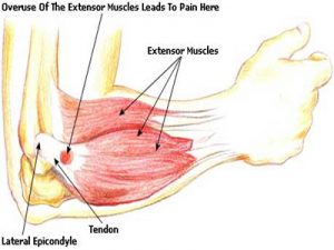
Tennis elbow, or lateral epicondylitis, is a painful condition of the elbow usually caused by overuse. Muscles, ligaments, and tendons support the functioning of the elbow joint. Your forearm extensor muscles and tendons extend the wrist and fingers. These extensor tendons attach the forearm muscles on the outside of the elbow to the lateral epicondyle. Tennis elbow is an injury to these tendons leading to pain and tenderness on the outside of the elbow. The main tendon involved in tennis elbow is usually the ”extensor carpi radialis brevis” tendon . Tennis elbow misdiagnosis is relatively common.

Tennis Elbow

When the tendon is weakened from overuse or trauma , microscopic tears form in the where the tendon attaches to the lateral epicondyle. This leads to pain and inflammation. The extensor carpi radialis brevis tendon may also be at increased risk of damage due to its position. As the elbow bends and straightens, the tendon rubs against the bone. This repetitive action can cause gradual wear and tear on the tendon over time in certain activities.

Activities – cause

Athletes are not the only people who get tennis elbow. Many people with tennis elbow participate in work or recreational activities that require repetitive and vigorous use of the forearm muscle. Painters, plumbers, carpenters, hairdressers, barbers, butchers(boners), people cutting hedges with clippers for long periods, are all particularly prone to developing tennis elbow.

Misdiagnosis

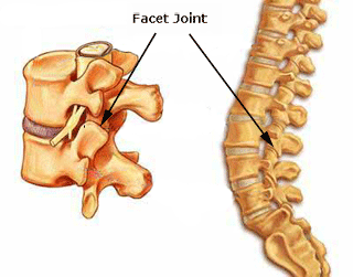
Tennis elbow misdiagnosis and mismanagement of this condition is often why people fail to recover. There are two common areas of misdiagnosis. Firstly if a person has not been doing any repetitive work with their forearm and there has been no forceful trauma while using the forearm, it is unlikely that the condition is tennis elbow. Sometimes the actual issue here is  nerve pain radiating down the arm from nerve compression in the neck and/or shoulder(neuropraxia). This nerve compression can be due to tight muscles/ locked facet joints and/or misaligned structures, often due to poor posture.  In the second type of tennis elbow misdiagnosis there can be repetitive injury involved but the condition is being exacerbated by nerve compression in the neck and shoulders, so both all areas need to be looked at for full resolution.

A cortisone injection is often used to try and treat ”tennis elbow”. This at most usually only gives short term relief and can even exacerbate the condition long-term with true tennis elbow, as it often allows the person to return to the aggravating activity thinking the injury has healed. Cortisone is an anti-inflammatory and may decrease inflammation in the area, reducing pain. It does not however stimulate tendon repair, which is what has been damaged in true ”tennis elbow” . Neither does cortisone do anything for a trapped nerve.

The above conditions are where an experienced physiotherapist/physical therapist can help greatly, usually much more so than any medication. This is their area of expertise.  However, you always need to remember accurate diagnosis and effective treatment is very practitioner dependent. Eccentric training along with soft tissue work can be very effective for true ”tennis elbow”. For the nerve compression (neuropraxia) described above, soft tissue release of neck and shoulder muscles(levator scapulae, trapezius,scalenes etc.) along with osteopathic manipulations to release any locked cervical or thoracic facet joints can be very effective. Symptoms of nerve entrapment/involvement can include neck/shoulder pain with tingling down the arms and/or into fingers. There are also manual orthopedic tests to check for nerve entrapment.

Aside : Carpal Tunnel syndrome is a medical condition due to compression of the median nerve as it travels through the wrist at the carpal tunnel. This can also be misdiagnosed and be due to nerve compression in neck /shoulder or upper thoracic regions.

Physiotherapists in Tralee  phone 086-7700191

Muscle twitches

Muscle TwitchesMuscles are made up of fibers that your nerves control. Muscle twitching is caused by minor muscle contraction in the area, or uncontrollable twitching of a muscle group that is served by a single motor nerve fiber. Stimulation or damage to a nerve may cause your muscle fibers to twitch. Most muscle twitches are minor and aren’t usually a cause for concern. Some are common and normal. Others are signs of a nervous system disorder.


Causes

These may include:

  • Autoimmune disorders such as Isaac syndrome.
  • Drug overdose (caffeine, amphetamines, or other stimulants).
  • Lack of sleep.
  • Drug side effect (such as from diuretics, corticosteroids, or estrogens).
  • Exercise (twitching is seen after exercise).
  • Lack of nutrients in the diet (deficiency).
  • Stress.
  • Medical conditions that cause metabolic disorders, including low potassium, and kidney disease, and uremia.
  • Twitches not caused by disease or disorders (benign twitches), often affecting the eyelids, calf, or thumb. These twitches are normal and quite common, and are often triggered by stress or anxiety . These twitches can come and go, and usually do not last for more than a few days.

Nervous system conditions that can cause muscle twitching include:

  • Amyotrophic lateral sclerosis (Lou Gehrig disease)
  • Neuropathy or damage to the nerve that leads to a muscle
  • Spinal muscular atrophy
  • Weak muscles (myopathy)


When to Contact a Medical Professional

Call your health care provider if you have long-term or persistent muscle twitches or if twitching occurs with weakness or loss of muscle.

Physiotherapist in Tralee : Phone 086-7700191

Carpal tunnel syndrome
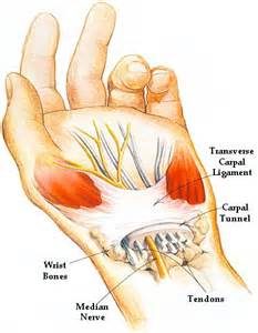
Carpal tunnel syndrome is a condition that causes numbness, tingling and weakness in the hand and arm. The condition is caused by  compression of the median nerve within the carpal tunnel, a narrow passageway on the palm side of your wrist. The median nerve runs from your neck  down along the arm and through the carpal tunnel to your hand. It provides sensation to the palm side of your thumb and fingers, except the little finger. It also provides nerve signals to move the muscles around the base of your thumb .  Anything that squeezes or irritates the median nerve in the carpal tunnel space may lead to this condition. The anatomy of your wrist, health problems and possibly repetitive hand motions may also contribute.   A wrist fracture can narrow the carpal tunnel and irritate the nerve, as can the swelling and inflammation resulting from rheumatoid arthritis.

Proper treatment usually relieves the tingling and numbness and restores wrist and hand function. It is worth noting that carpal tunnel syndrome is often misdiagnosed and the main site of median nerve compression can be occurring up at neck and shoulder level. Often by freeing up neck and shoulders muscles and surrounding joints with physio as well as working out muscles down along the arm through which the median nerve penetrates, and loosening and stretching the wrist structures, much relief, often total relief can often be achieved.


Symptoms

Syndrome symptoms usually start gradually.

Numbness: The first symptoms are usually tingling or numbness usually in the thumb, index and middle or ring fingers, but not your little finger. The sensation may travel from your wrist up your arm. These symptoms often occur while holding a steering wheel, phone or newspaper. The sensation may wake you from your sleep.

Weakness: You may experience weakness in your hand and a tendency to drop objects. This may be due to the numbness in your hand or weakness of the thumb’s pinching muscles, which are also controlled by the median nerve.


Carpal Tunnel Risk factors

A number of risk factors have been associated with carpal tunnel syndrome.  Although they may not directly be the cause , they may increase your chances of developing or aggravating median nerve damage.

These include:

Anatomic factors: wrist fracture, dislocation, or arthritis that deforms the small bones in the wrist, can alter the space within the carpal tunnel and put pressure on the median nerve.

Sex: It is generally more common in women. This may be because the carpal tunnel area is relatively smaller in women than in men.

Nerve-damaging conditions: Some chronic illnesses, such as diabetes, increase your risk of nerve damage.

Inflammatory conditions: Illnesses that are characterized by inflammation, such as rheumatoid arthritis, can affect the lining around the tendons in your wrist and put pressure on your median nerve.

Obesity : Being obese is a significant risk factor for carpal tunnel syndrome.
Alterations in the balance of body fluids. Fluid retention may increase the pressure within your carpal tunnel, irritating the median nerve. This is common during pregnancy and menopause. Carpal tunnel syndrome associated with pregnancy generally resolves on its own after pregnancy.


Carpal Tunnel Diagnosis

History of symptoms, Physical examination. Nerve tension tests.

X-ray. Some doctors recommend an X-ray of the affected wrist to exclude other causes of wrist pain, such as arthritis or a fracture.

Electromyogram. This test measures the tiny electrical discharges produced in muscles. During this test, your doctor inserts a thin-needle electrode into specific muscles to evaluate the electrical activity when muscles contract and rest. This test can identify muscle damage and also may rule out other conditions.

Nerve conduction study. In a variation of electromyography, two electrodes are taped to your skin. A small shock is passed through the median nerve to see if electrical impulses are slowed in the carpal tunnel. This test may be used to diagnose your condition and rule out other conditions.


Treatment

If the condition is diagnosed early, nonsurgical methods may help improve carpal tunnel syndrome. Physical therapy by a practitioner experienced in this area can be very effective. This would most like involve deep tissue work, osteopathic manipulations, electroacupuncture and postural correction exercises, and nerve glide exercises as the condition improves. Other treatment options include wrist splinting, taking more frequent breaks to rest your hands, avoiding activities that worsen symptoms, medications(Nonsteroidal anti-inflammatory drugs) icing, rest etc. Surgery is also an option but is a last resort.


For treatment of this condition contact us on 0867700191


For more information see this video


Restless Legs Syndrome 

RESTLESS LEG SYNDROME
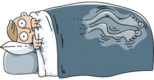
Restless legs syndrome(RLS) is a nervous system disorder resulting in an urge to move the legs. Because it usually interferes with sleep, it also is considered a sleep disorder. The condition causes an uncomfortable, “itchy,” “pins and needles,” or “creepy crawly” type feeling in the legs. These sensations are usually worse at rest, especially when lying or sitting. The symptoms are generally also worse in the evening and at night. The severity of RLS symptoms varies from mild to intolerable. Symptoms can come and go and vary widely in severity. For some people, symptoms may lead to severe sleep disturbance at night. This can significantly impair their quality of life.


Who Gets Restless Legs Syndrome?

It is estimated that ”Restless legs syndrome” (RLS) affects up to 10% of the population. It affects both sexes, but is more common in women. The condition may begin at any age, but most people affected are middle-aged or older. RLS is often unrecognized or misdiagnosed. This is especially true if the symptoms are intermittent or mild.


Causes of Restless Legs Syndrome

In most cases, doctors do not know the cause of restless legs syndrome. It is suspected that genetics plays a role. Nearly half of people with RLS also have a family member with the condition. Factors other than genetics are associated with the development of restless legs syndrome. Certain chronic diseases and medical conditions can cause RLS. These include iron deficiency, Parkinson’s disease, kidney failure, diabetes, and peripheral neuropathy. Treating these conditions often gives some relief from RLS symptoms.


Medications

Some types of medications may exacerbate symptoms. These include anti-nausea drugs, antipsychotic drugs, some antidepressants, cold and allergy medications containing sedating antihistamines. Women can experience RLS during pregnancy, especially in the last trimester. These symptoms usually go away within a month after delivery. Other factors, including alcohol use and sleep deprivation, may trigger symptoms or make them worse.


Diagnosis

There is no medical test to diagnose RLS. Doctors. However, blood tests or other diagnostic tests may be used to rule out other conditions. The diagnosis of RLS is based mainly on a patient’s symptoms and history .


Treatment

Treatment of RLS is targeted at easing the symptoms. In people with mild to moderate restless legs syndrome, lifestyle changes, such as beginning a regular exercise program , establishing regular sleep patterns, and eliminating or decreasing the use of caffeine,alcohol and tobacco, may be helpful.

Non-drug RLS treatments may include:

Leg massages

Hot baths or heating pads or ice packs applied to the legs

Good sleep habits

A vibrating pad called Relaxis


Drugs

Medications may sometimes be helpful in treatment of RLS but results vary among individuals. Drugs used to treat RLS include:

Dopaminergic drugs, which act on the neurotransmitter dopamine in the brain; Mirapex, Neupro, and Requip, levodopa.

Benzodiazepines, a class of sedative medications, may be used to help with sleep, but they can cause daytime drowsiness.

Narcotic pain relievers may be used for severe pain.

Anticonvulsant drugs  such as Tegretol, Lyrica, Neurontin,  and Horizant.


Although there is no cure for restless legs syndrome, current treatments can help control the condition, decrease symptoms, and improve sleep.


For more information see these videos     video 1.           video 2.


Restless legs syndrome treatment  by physical therapist in Tralee, Co. Kerry. Phone Eddie on 086-7700191. Click for Website

physiotherapist
Fig. 1. pronation-supination

Physiotherapist explains foot pronation & supination

A physiotherapist explains Pronation and supination as  movements that occur at the subtalar joint of the foot. The normal biomechanics of the foot are designed to absorb and direct the forces occurring throughout the gait cycle. As the foot is loaded, eversion of the subtalar joint, dorsiflextion of the ankle, and abduction of the forefoot occur. This is the pronation part of the gait cycle. Pronation should not continue past the latter stages of midstance during the gait cycle. At this stage the foot should then supinate in preparation for toe-off. Approximately four degrees of pronation and supination are necessary to enable the foot to propel forward properly. Any increase on this four degrees brings a foot into over-supination or over-pronation.


Pronation – physiotherapist explaination

Pronation of the foot is where the heel and the little toe move away from the center of the body. The foot also dorsiflexes up slightly, the ankle rolling inwards. Pronation is part of the natural movement of the human body. Certain injuries can occur with excessive pronation.  Runners with flat feet often tend to overpronate. Over-pronation can contribute to many injuries. These include shin splints, anterior compartment syndrome, patello-femoral pain syndrome, plantar fasciitis, tarsal tunnel syndrome, bunions (hallux valgus), achilles tendonopathies etc. The running shoes of over-pronators often show extra wear on the inner heel and ball of the foot.


Supination – physiotheraist explaination

With supination the heel and also the big toe rotate towards the centre of the body. The foot flexes down and the ankle rolls out. It is the opposite of pronation. A natural amount of supination occurs during the push-off phase of the running gait. This occurs as the heel lifts off the ground and the forefoot and toes are used to propel the body forward. However, excessive supination  places a large strain on the muscles and tendons that stabilize the ankle. This can make the oversupinator more prone to ankle sprain or ankle ligament rupture.

With over-supination the forces of impact on the foot are concentrated on a smaller area of the foot (the outside part), and are not distributed as efficiently. In the push-off phase, most of the work is done by the smaller toes on the outside of the foot, rather than the big toe. This places extra stress on the foot. It can lead to conditions such as iliotibial band syndrome, Achilles tendinitis, or plantar fasciitis. Over-supination causes the outer edge of running shoes to wear sooner. In extreme cases, there will also be holes in the uppers where the runner’s foot has broken through. Runners with high arches and tight Achilles tendons/calves tend to be over-supinators.


Orthotics

Properly prescribed foot orthotics can be beneficial in the treatment of over-pronation or over-supination. Physiotherapists usually supply both off the shelf and/or custom made orthotics. We  prescribe these only when we really feel they are necessary. Often the much cheaper off the shelf version will do the trick. It is important to get a quality product, prescribed by an experienced practitioner. Sometimes you just have an injury that needs  treatment and there is no need for an orthotic. Also high arches or a flat feet do not mean in itself you need orthotics, especially if you are not in pain.


Bone Spurs – Causes?

Bone spurs(also called osteophytes) are outgrowths of bone occurring along the edge of a bone. Bone spurs are most commonly found in joints, where two bones come together. They also occur close to where muscles, ligaments, or tendons attached to bone. Some common parts of the body affected include the neck (cervical spine), shoulders, hips, hands and heel bone.

Bone spurs typically occur because of continued stress or rubbing on a bone over a prolonged period of time. This can occur due to inflammatory conditions  such as osteoarthritis , tendinitis or tenosynovitis. Normally there is a smooth layer of cartilage on the edges of bones where they come together to form a joint. With osteoarthritis this cartilage layer becomes worn away. This causes the exposed bones to rub against each other. New bone forms in response to this stress and the resulting inflammation. Bone spurs usually develop in areas near tendons and ligaments due to chronic inflammation in these areas. This inflammation can result from friction between these tissues and bone, or from overuse. The bone spur development is the bodies way of trying to protect itself.

Signs and Symptoms

Bone Spurs
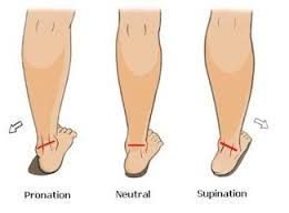
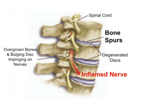
Bone spurs may or may not cause symptoms. Symptoms are location dependent. If bone spurs rub against other bones at joints they can cause pain and/or more limited movement in these joints. They can also be associated with pain, numbness, tenderness, and /or weakness in areas where  they irritate adjacent tissue structures(muscles, ligaments, bones, nerves etc.). If the bone spur rubs against tendons or ligaments, they can sometimes cause a tear in these tissues over time. This is a common cause of tears in certain rotator cuff tendons .

If bone spurs occur in the spine  they can sometimes pinch the nerves (radiculopathy) or spinal cord(myelopathy). A radiculopathy can cause pain, numbness, tingling, or weakness in the arms or legs depending on the area affected. With a myelopathy there can be pain and problems with balance along with weakness.

Diagnosis

Generally X-Rays, MRIs or CT scans are used to confirm suspected cases. Sometimes an experienced practitioner can feel the spur with their finger depending on the area(i.e. heel spur).

When Is Surgery Appropriate ?

In some cases, if symptoms cannot be controlled with more conservative treatment, surgery could be an option. The goal is to remove the bone spur, allowing a return to normal joint motion, or to remove the pressure on muscles, tendons, ligaments, or nerves.


Physiotherapist Tralee  :  Phone 086-7700191

Dead leg

Dead Leg
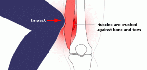
A ”dead leg”, also known as ”charley horse” or ”quadriceps contusion”, is essentially an injury due to a traumatic blow, crushing the quadriceps muscle against the femur bone. The quadriceps is the muscle at the front of your thigh. The injury can be either intermuscular or intramuscular. Treatment depends on the type of contusion and grade in severity of the injury. An Intramuscular contusion occurs when the muscle gets torn within the sheath surrounding it. This causes the initial bleeding to cease within hours due to increased pressure within the muscle. However, the fluid and blood is not able to escape from the muscle sheath surrounding it resulting in considerable loss of function and a lot of pain. This can take days or weeks for a full  recovery. You are unlikely to see any bruising with this type of contusion, especially in the early stages. In the case of intermuscular contusions, the muscle as well as part of the sheath surrounding it gets torn. This results in a longer bleeding time initially, especially if there is no use of ice therapy. The patient usually recovers faster from this type of dead leg, as the blood and fluids can easily flow away from the injury site. Bruising is often present in this type of contusion.

Grading

  • Grade 1: Quadriceps pain, tightness in the thigh and a limp while walking. The swelling is very mild and so is the pain. The patient has almost complete range of motion upon stretching.
  • Grade 2: The patient is not able to walk properly and limps when walking. There is some swelling present and the patient will experience bouts of pain upon activity. There is pain upon extending the leg against resistance. Pressure on the site also produces pain and the patient is unable to completely flex the knee.
  • Grade 3:The patient has severe pain with obvious swelling, and is unable to walk without the help of crutches. Muscle contraction produces an obvious gap or bulge. This type of dead leg needs at least a month or two to completely heal.

Treatment

  • I.C.E (Rest, Ice, Compression, Elevation).
  • Rest is very important and the limb should be kept elevated as much as possible.
  • Compression and ice therapy should be applied for 15 to 20 minutes every hour for the first 24 to 48 hours. Ice should be wrapped in a wet towel to prevent ice burns. A compression bandage should be worn 24/7 to reduce swelling and to give support until the pain has subsided.
  • Crutches should be used if required.
  • Never apply heat therapy(that includes having a hot bath…Do not) or perform deep tissue massage in the initial acute stage (24 to 48 hours) of a dead leg injury. This can aggravate the injury or lead to myositis ossificans later. Sports massage to help recovery should be started once the acute phase (24 to 48 hours) of the injury has passed, provided it’s not an intramuscular contusion(This may need more time to settle and sometimes requires medical intervention).
  • Gentle stretching of the area should be done after the acute stage (24 to 48 hours) has passed, but not before, provided it does not cause too much pain. The stretch should be held for 30 seconds and should be repeated 4 to 5 times in a day. With severe (grade 3) intermuscular contusion there should be no overpressure with passive stretching for 7-10 days.


When Can Exercises Be Started?

  • For dead leg of grade 1 contusion of quadriceps, strengthening exercises can be started as soon as the pain has subsided. Patient should start with mild exercises and should move on to difficult ones as the dead leg pain.
  • For dead leg of grade 2 contusions of quadriceps, strengthening exercises should be started after a week or so. Patient should start with gentle warm up exercises and finish with good stretching exercises. Strengthening exercises should be continued for several weeks. Strengthening exercises should be started more than a week later and for grade 3 contusions of the quadriceps.
  • Gentle exercises such as cycling or swimming are nice gentle strengthening exercises to start with.


Myositis Ossificans

The more severe a contusion, the greater the risk of development of Myositis Ossificans, especially with poor treatment and management. In this condition calcification occurs within the healing hematoma(swelling of clotted blood within the muscle). The healing hematoma forms bone within the layers of affected muscle. Symptoms include overnight and morning pain, as well as pain on muscle contraction. It may be possible to feel a hard bump or ‘woody’ lump within the affected muscle tissue.  Stiffness and loss of knee range of movement are also common.  Sometimes surgery is required to remove the calcified tissue.

Physiotherapists in Tralee

Back Pain Treatment Options Made Simple

Three very common causes of back pain presenting in clinic are 1. muscular, 2. discogenic (from a disc), 3. locked facet joint. We will look at each and the relevant back pain treatment options.

Muscular back pain

For the low back a common muscle to cause trouble is the piriformis muscle (see diagram). It is involved in hip rotation movements so if it tightens you lose rotation putting pressure on the lower back during activities. When the piriformis muscle tightens it may also squeeze on the sciatic nerve which travels from the lumbar spine  down the back of the leg causing pain anywhere along the nerve path(low back, hamstring, calf, sole of foot). Tightened hamstrings may also contribute to low back pain(often resulting from being seated for long periods) making the spine do more work due to resulting poor posture in lifting etc.

The piriformis muscle can usually be worked out and loosened  with deep tissue massage.  The hamstrings can also be worked if these are contributing. Electro-acupuncture also helps loosen the muscles/trigger points and settle nerve irritation in affected areas.

Back pain

Locked Facet joint back pain

This is a common cause of back pain in the upper thoracic region(along back of the rib cage)…Often the person feels like somebody is sticking a finger into their back when they inhale deeply. Also with this condition a person may find that they can turn completely in one direction but not the other. In more serious cases pain may shoot down the arm due to nerve entrapment/irritation. A locked facet joint may also occur in the lower back sending nerve pain down the leg. The crack you hear when the back is manipulated is locked facet joints opening, it is not a ”slipped disc” being put back into place. A timely spinal manipulation done correctly, with a little soft tissue work done thereafter can often sort this condition. With the manipulation you get a little crack if the facet joint was locked, as it opens, giving immediate relief.

Back pain


Discogenic (Disc) related back pain

This is a more tricky one. Probably most common in the lower back (lumbar region). The patient will sometimes present with their spine all twisted off to one side in a sort of ”S” shape. This is usually because the disc bulge or protrusion is touching a nerve. The body compensates for this by trying to get the disc away from the nerve, hence the ”S” shape. Also there will be muscle spasm as the body tries to protect itself. A patient with this condition is usually in a lot of pain and even simple tasks like walking can be excruciatingly painful. Disc pain is less common in the thoracic region but again here it presents with deep unrelenting pain.

There is no such thing as a ”slipped disc”, it is just a misnomer. Discs basically bulge, degenerate(collapse) or herniate(a little fluid filled sack can protrude or leak out of them)(see diagram below). It is when this bulge or protrusion touches a nerve, the trouble starts.

Basically a muscle relaxant (valium) from your doctor along with an anti-inflammatory (NSAIDS) for a couple of days usually helps a lot.  Usually two treatment sessions along with the medication to loosen out the muscles will also reduce pain and improve recovery time. A manipulation to open any locked facet joints may help as the condition settles.

When a disc touches a nerve it initiates a series of reactions including muscle spasm and inflammation. The medication is very important in this condition and the combination of the both muscle relaxant and anti-inflammatory seem to complement each other along with treatment. Things such as ”Mckenzie technique” and ”nerve glides” can also help when introduced at the right time during treatment but require too much detail to go into here.

Once the condition settles it is important to 1. Train your body to engage its core. 2. Strengthen your core muscles with pilates type exercises, along with developing your core fitness. 3. Lose weight if you are overweight. 4. Try and improve your flexibity.

Once you get somebody moving, loosened out, give them a proper rehab routine and they are taking the medication, it is more about giving the body time to heal itself than anything else. Basically what you are trying to do with initial treatment is to relax any muscle spasm, reduce inflammation, reduce pressure on the disc with the more long-term goal of strengthening the body in the hope of bringing back in the disc bulge/ protusion enough from the nerve it is touching, so that symptoms subside. If there is little improvement in your condition after a week, an mri may be required to get more accurate information on the injury.  In some cases an epidural or surgery  may be required.

Back pain

Please note the above article only lists some common causes of back pain.

Back pain treatment in Kerry –  phone 086-7700191


Metatarsalgia

Metatarsalgia
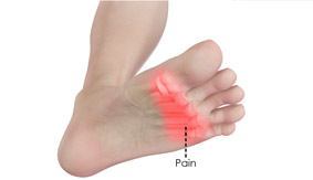
Metatarsalgia is the name given to pain in the front part of your foot under the heads of your metatarsal bones ( ball of foot, just before toes). It is usually worse when standing or walking etc.  and occurs most frequently in the second, third/or fourth metatarsal joints or isolated in the first metatarsal joint. Metatarsalgia usually comes on gradually over some weeks rather than suddenly. The affected area of your foot may also feel tender on palpation by your physiotherapist.

Common causes identified by physiotherapists include:

  • Pes cavus or high arched foot.
  • Excessive pronation of the foot(foot rools inwards ie. with flat foot).
  • Clawing or hammer toes.
  • Tight extensor tendons of the toes.
  • Prominent metatarsal heads.
  • Morton’s foot – here there is a shortened first metatarsal, which results in an abnormal gait putting increased pressure on the second metarsal.
  • Over doing it in athletes such as runners and tennis players etc. can lead to inflammation in the joints due to the pounding they receive.
  • Being overweight puts extra stress on many areas of the foot including metatarsals.
  • Wearing high heels – forward force on feet increases the pressure on metatarsal area.
  • Having tightened calf muscles or poor ankle flexibility – this affects the normal gait pattern.
  • Poorly fitting or tight footwear can squeeze the metatarsal joints causing friction , inflammation and poor gait.

Simple measures can help to relieve the symptoms of metatarsalgia. These include:

  • Resting with your feet elevated where possible.
  • Losing weight if you are overweight.
  • Wearing supportive shoes that are well fitted, low-heeled and have a wide toe area.
  • Metatarsal pads and orthotic inserts for your shoes may help to relieve pain in your forefoot by reducing the pressure placed on the heads of your metatarsal bones.
  • Physiotherapy may also be helpful. This may include stretching out the spaces between the metatarsal and mobilising the joints of the foot or performing deep tissue massage to loosen your calf muscles or any other lower limb muscles that may be contributing to poor gait. Sometimes if a metatarsal head has dropped it can be manipulated back into place. Pain relief as a result of this mobilisation, when effective can be immense and long lasting.
  • Simple painkillers such as paracetamol and non-steroidal anti-inflammatories(NSAIDs) such as ibuprofen may help to relieve pain.