spinal manipulationSpinal manipulation is  a technique where practitioners use their hands or a device to apply a controlled thrust to a joint of your spine. The amount of force can vary, but the thrust moves the joint more than it would on its own. It is designed to relieve pressure on joints, reduce inflammation, and improve nerve function. It’s often used to treat back, neck, shoulder, and headache pain in combination with other treatments i.e. deep tissue massage, electrotherapy, rehabilitation exercises. The common goal of most spinal manipulations is to restore or to enhance joint function, with the general goals of resolving joint inflammation and reducing pain.

Spinal Manipulation Techniques

Physiotherapists and osteopaths adapt treatment plans to meet the specific needs of each patient. Some of these treatment plans involve some forceful and less forceful spinal adjustment techniques during the same visit or over the course of treatment .

Spinal Manipulation (High-Velocity Low-Amplitude Thrust)

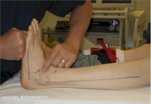
The most frequently used technique, spinal manipulation, is the traditional high-velocity low-amplitude (HVLA) thrust. The manipulation often results in an audible “pop,” as physiotherapists use their hands to apply a controlled sudden force to a joint while the body is positioned in    a specific way.


Is the audible pop necessary ?

The sound often heard during an HVLA manipulation is called cavitation. The pop is caused by a release of gas when the joint is pushed a short distance past its passive range of motion of the joint. The mechanism is similar to cracking ones knuckles.  An audible pop isn’t always necessary for the treatment to be successful, but everybody seems happier when there is one.


Spinal Mobilization (Low-Force / Gentle Techniques)

Some patients and/or clinicians prefer mild spinal mobilization techniques that do not involve twisting of the body or a forceful thrust.The goal of spinal mobilization is the same as HVLA spinal manipulation – to restore or to enhance joint function. However, unlike HVLA spinal manipulation, slow movement, usually to a firm endpoint of joint movement, is used to mobilize the joint.

Physiotherapists may choose spinal mobilization for certain patients for a variety of reasons, such as:

  • Patient preference – certain patients prefer spinal mobilization over spinal manipulation.
  • Obesity can make the positioning of the patient and the manipulation procedures challenging for both the provider as well as the patient, which might favor a low force technique.
  • Patients with sensitive nervous systems may benefit from gentle mobilization techniques to keep the body from overreacting and causing reactive muscle spasms.
  • Patients with some conditions may be contraindicated for spinal manipulation, such as possibly patients with advanced osteoporosis, bone pathology, some forms of deformity, and certain types of inflammatory arthritis.
  • Physiotherapists may choose spinal mobilization for patients when they are in the acute stage of their condition and in severe pain.
  • Obesity can make the positioning of the patient and the manipulation procedures challenging for both the provider as well as the patient, which might favor a low force technique.


See video below demonstrating spinal manipulation

https://youtu.be/mxzHUYF6LBQ


Osgood Schlatter disease : Knee Pain in Adolescents

Osgood Schlatter disease is an inflammation of the bone, cartilage and/or tendon at the top of the tibia (shinbone ie. just under the knee), where the tendon from the kneecap(patella) attaches. It presents as a painful lump below the kneecap. It usually strikes active adolescents during growth spurts which can begin any time between the ages of 8 and 13 in girls, 10 and 15 years  in boys, basically during puberty.  Growth spurts make kids vulnerable because their bones, muscles, and tendons are growing quickly and not always at the same time. With exercise, these differences in size and strength between the muscle groups place unusual stress on the growth plate at the top of the shinbone (A growth plate is a layer of cartilage near the end of a bone where most of the bone’s growth occurs. It is weaker and more vulnerable to injury than the rest of the bone).


While  Osgood Schlatter disease  is more common in boys, the gender gap is narrowing as more girls become involved in sports.  Osgood Schlatter disease  affects as many as 1 in 5 adolescent athletes. Teens increase their risk if they play sports involving running, twisting, and jumping, such as basketball, football, volleyball, soccer, tennis, figure skating, and gymnastics etc. Doctors disagree about the mechanics that cause the injury but agree that both overuse and physical stress are involved. Most parents call the doctor after their child complains of intermittent pain over several months.

Signs and symptoms of  Osgood Schlatter disease

  • pain that worsens with exercise
  • relief  from pain with rest
  • swelling or tenderness at the bony prominence under the knee and over the shinbone
  • limping after exercise
  • tightness of the muscles surrounding the knee (the hamstring sometimes but particularly the quadriceps muscles)


The pain varies from person to person. Some have only mild pain while performing certain activities. For others, the pain is nearly constant and can be debilitating. The good news is that Osgood Schlatter disease (OSD) is far less serious than its name suggests. Though it is one of the most common causes of knee pain in adolescents, it is really not a disease, but an overuse injury. The condition is self limiting and usually resolves itself within 12 to 24 months. A similar condition, ”Sever’s disease” occurs in children of the same age group down at the achilles tendon attachment and presents as heel pain. Again the condition is self limiting and the same treatment protocols apply.

While there is no evidence that rest accelerates the healing process, a reduction in activity usually reduces the pain. There is no need to rest completely. Pain should be the main guide as to limitation of activity. Tightness in  the quadriceps muscles may predispose to this condition. The athlete should commence a stretching program. Some massage therapy on the quadriceps  can be tried. Pain also can sometimes be relieved by icing the painful area(ice in wet towel) for 10 minutes at a time. Correction of any biomechanical abnormality, such as excessive subtalar pronation(feet turn out while running) should also be considered.

Summary

To sum up, this is basically a self limiting condition that adolescents generally grow out of (Brukner and Khan, 2011). Some stretching exercises  and modification of activities is generally the best and most cost effective course of treatment.

References

Brukner P, Khan K. Clinical Sports Medicine. 4th ed. Sydney: McGraw-Hill, 897-898, 2011


For more information or to make an appointment phone 086-7700191. Physiotherapists in Tralee, Co. Kerry.

Bunions

Bunions develop slowly. The big toe is made up of two joints. A bunion is a painful bony bump, also known as hallux valgus that develops on the inside of the foot at the big toe joint.  Pressure on the big toe joint causes it to bend inwards towards the second toe. Over time, the normal structure of the joint changes, resulting in a bunion. The enlarged joint is often inflamed. The deformity gradually increases making it painful to wear shoes or walk. Bunions are more common in women. Many women wear tight, narrow shoes that squeeze the toes together. This makes it more likely for a bunion to develop. In most cases, bunion pain is relieved by wearing wider shoes with adequate toe room and using other simple treatments to reduce pressure on the big toe.

Bunion Progression

An advanced bunion can greatly alter the appearance of the foot. In severe bunions, the big toe may angle all the way under or over the second toe. Pressure from the big toe may force the second toe out of alignment, causing it to come in contact with the third toe. Calluses may develop where the toes rub against each other, causing additional discomfort and difficulty walking.

Foot Problems Related to Bunions

In some cases, an enlarged big toe joint may lead to bursitis, a painful condition in which the fluid-filled sac (bursa) that cushions the bone near the joint becomes inflamed. It may also lead to chronic pain and arthritis if the smooth articular cartilage that covers the joint becomes damaged from the joint not gliding smoothly.

 Causes

Bunions may be caused by:

  • Wearing poorly fitting shoes—in particular, shoes with a narrow, pointed toe box that forces the toes into an unnatural position
  • Heredity—some people inherit feet that are more likely to develop bunions due to their shape and structure
  • Having an inflammatory condition, such as rheumatoid arthritis, or a neuromuscular condition, such as polio.

Diagnosis of bunions

Physical examination of bunions

Your doctor will ask you about your medical history, general health, and symptoms. He or she will perform a careful examination of your foot. Although your doctor will probably be able to diagnose your bunion based on your symptoms and on the appearance of your toe, he or she will also order an x-ray.

X-Rays

X-rays provide images of dense structures, such as bone. An x-ray will allow your doctor to check the alignment of your toes and look for damage to the MTP joint.

Nonsurgical treatment of bunions

In most cases, bunions are treated without surgery. Although nonsurgical treatment cannot actually “reverse” a bunion, it can help reduce pain and keep the bunion from worsening.

Changes in Footwear

In the vast majority of cases, bunion pain can be managed successfully by switching to shoes that fit properly and do not compress the toes.

Padding

Protective “bunion-shield” pads can help cushion the painful area over the bunion. Pads can be purchased at a drugstore or pharmacy. Be sure to test the pads for a short time period first; the size of the pad may increase the pressure on the bump. This could worsen your pain rather than reduce it.

Orthotics and Other Devices

Orthotics (custom-made shoe inserts) may be used to take pressure off your bunion. Toe spacers can also be placed between your toes to try and straighten the big toe. In some cases, a splint worn at night that places your big toe in a straighter position may help relieve pain.

Icing

Applying ice several times a day for 20 minutes at a time can help reduce swelling. Do not apply ice directly on your skin.

Medications

Nonsteroidal anti-inflammatory medications such as ibuprofen  can help relieve pain and reduce swelling. Other medications can be prescribed to help pain and swelling in patients whose bunions are caused by arthritis.

Bunions and surgery

Your doctor may recommend surgery for a bunion, after a period of time, if you still have pain and difficulty walking despite changes in footwear and other nonsurgical treatments. Bunion surgery realigns bone, ligaments, tendons, and nerves so that the big toe can be brought back to its correct position.


Physiotherapists in Tralee stocking a wide range of orthotics to treat various foot conditions. Phone 0867700191


equinus

Equinus Foot Condition

The ankle joint connects the leg to the foot. It is formed by three separate bones, the tibia, fibula and talus. The shinbone (tibia) supports most of a person’s weight when standing. The outer bone (fibula) is the smaller bone of the lower leg. A small, irregular-shaped foot bone (talus) connects the tibia and fibula. Acting as a hinge, these bones form the ankle. The ankle joint allows movement such as walking, running and jumping, and also contributes to lower limb stability.

The ankle is reinforced by ligaments which connect bone to bone. Ligaments have a mildly elastic structure that allows them to stretch, within their limits, and then return to their normal positions. Ligaments protect the ankle from abnormal movements—especially twisting, turning and rolling of the foot.

Description

A person with equinus has a limited range of ankle motion and lacks the flexibility needed to bring the top of the foot upward, toward the shin. It may be either congenital or acquired, and occurs equally in both men and women.  Equinus can be due to several different reasons including the following:

• bony block between the talus and distal tibia (osseous equinus);
• contracture or tightness of the soleus muscle (inner calf muscle);
• contracture or tightness of the soleus and gastrocnemius muscles (gastroc-soleal equinus);
• isolated tightness of the gastrocnemius muscles (outer calf muscle); and
• compensatory loss of ankle joint range of motion for some other condition such as pes cavus (pseudoequinus).


People with equinus often develop ways to compensate for their limited ankle motion. Depending on how a patient compensates for the inability to bend properly at the ankle, a variety of other foot conditions can develop, such as:

  • Plantar Fasciitis
  • Calf cramping
  • Achilles Tendinitis
  • Metatarsalgia (pain and/or callusing on the ball of the foot)
  • Flatfoot
  • Arthritis of the midfoot (middle area of the foot)
  • Pressure sores on the ball of the foot or the arch
  • Bunions and hammertoes
  • Ankle pain
  • Shin splints
  • Sesamoiditis
  • Hallux valgus
  • hallux rigidus
  • Hammer toes


Nonsurgical Treatment of Equinus

Some nonsurgical treatment strategies are aimed at relieving the symptoms and conditions associated with equinus. Treatment for the equinus itself may include one or more of the following options.

  • Heel lifts—Placing heel lifts inside the shoes or wearing shoes with a moderate heel may reduce symptoms by taking stress off the Achilles tendon and compensating for the restricted movement of the ankle joint. The joint is meant to have about fourteen degrees of movement ideally, so say it has only four degrees of movement, adding a ten degree heel lift helps compensate for the missing degrees of movement .
  • Arch supports or orthotic devices—Custom orthotic devices that fit into the shoe are often prescribed to ensure that weight is distributed properly, and to help control muscle/tendon imbalance. Again these devices will most likely include a heel lift.
  • Physical therapy—To help remedy muscle tightness, deep tissue massage of calf muscles along with a stretching program for the calf muscles are recommended.


For a quick simple video describing the condition click   here


See our wide range of orthotics  here


What is Hallux Rigidus?

Hallux Rigidus

Big toe joint arthritis (otherwise known as Hallux Rigidus) is a form of degenerative arthritis. In this condition, surfaces of the joints in the big toe begin to wear away and extra bone can also develop in the form of  bone spurs or osteophytes. This limits the movement of the joint. The big toe  needs to bend significantly when stepping off. Consequently, arthritis in this joint can greatly affect walking, running etc.

Hallux Rigidus is a condition that tends to get worse over time. In it’s earlier stages, it may be referred to as Hallux Limitus, which is where there is limited movement of the big toe joint. With progression, Hallux Rigidus develops which can result in stiffness developing in the big toe joint and there may also be swelling.

To compensate for pain in the big toe joint, people with Hallux Rigidus tend to adjust their walking pattern which can also result in knee, hip or lower back pain. There is often difficulty finding shoes that fit properly shoes due to pain and/or inflammation in the joint. Early diagnosis can be made by physical examination and x-ray. Early treatment gives the best chance of avoiding surgery.

What causes Hallux Rigidus?

The big toe joint is designed to bear a considerable amount of stress during walking. However faulty biomechanics or structural abnormalities such as flat feet can stop the big toe from bending normally, and eventually lead to the development of osteoarthritis in the big toe joint. Other factors that can increase the risk of developing this condition include genetics (eg, having a certain foot type), injury to the big toe , other inflammatory diseases, and working in a job that places excessive stress on the big toe joint .

Treatment

In cases of Hallux Rigidus, certain types of footwear and orthotics may be recommended to reduce pressure and motion on the big toe.  Custom orthotics that conform very closely to the arch of the foot are most effective in improving big toe joint function. These orthotics may incorporate  something like a 3mm kinetic wedge in combination with a built in forefoot posting to take the pressure off the big toe joint. Ice and anti-inflammatory medication can also be used to reduce inflammation and pain. If it does not respond to conservative treatment, surgery may be recommended as a last resort in more severe cases.

Posterior ankle impingement physiotherapyPosterior ankle impingement is a condition characterised by tissue damage at the back of
the ankle joint due to compression of these structures. This occurs when the foot and ankle
are pointed maximally away from the body (plantarflexion – figure 1. ). It may occur when
compressive forces are too repetitive and/or too forceful. This can occurs in the presence of
ankle swelling or bony anomalies, such as additional bone, a condition known as an “os
trigonum”. Posterior ankle impingement is most commonly found in gymnasts, ballet
dancers, and footballers, because they regularly maximally plantarflex their ankles during
their activities. The condition can also occur due to inadequate rehabilitation of an acute
ankle injury (ie. ankle sprain).

Mechanism of Injury

Posterior ankle impingement may develop due to an acute traumatic plantar hyperflexion
event, such as an ankle sprain. It may also occur as a result of repetitive low-grade trauma
associated with plantar hyperflexion, say like in case of a female ballet dancer. It is
important to differentiate between these two, because the latter, that is posterior
impingement from overuse, has a better prognosis.
The anatomy of the posterior ankle is a key factor in the occurrence of posterior
impingement syndrome . The more common causes of the condition are osseous in nature,
such as the os trigonum, an elongated posterolateral tubercle of the talus (known as
Stieda’s process), a downward sloping posterior lip of the tibia, an osteophyte from the
posterior distal tibia , or a prominent posterior process of the calcaneus. However, posterior
impingement can also be soft tissue related, as with a thickened posterior joint capsule ,
post-traumatic scar tissue, post-traumatic calcifications of the posterior joint capsule, or
loose bodies in the posterior part of the ankle joint. Symptoms for all of these conditions
relate to physical impingement of osseous or soft tissue structures, resulting in painful
limitation of the full range of ankle movement.
The most common cause ''os trigonum'' is an extra (accessory) bone that sometimes
develops behind the ankle bone (talus). The mineralized os trigonum appears between the
ages of 7 and 13 years and usually fuses with the talus within 1 year, forming the trigonal
(Stieda) process. It may remain as a separate ossicle in 7-14% of patients, and is often
bilateral(in both ankles). An os trigonum can be a focus of osseous abutment against other
structures. Pain can also be caused by disruption of the cartilaginous synchondrosis
between the os trigonum and the lateral talar tubercle as a result of repetitive microtrauma
and chronic inflammation.
In the case of soft tissue impingement it usually results from scarring and fibrosis associated
with synovial, capsular, or ligamentous injury ie. bad ankle sprain. It is thought that this
type of manifestation usually usually occurs when a significant soft-tissue component
forms. The soft-tissue component can consist of synovial thickening throughout the
posterior capsule or be more focal, involving the posterior intermalleolar or talofibular ligament. The flexor hallucis longus tendon runs in the groove between the lateral and
medial processes of the talus and can also be injured in posterior impingement, resulting in
tenosynovitis.


Signs and symptoms

Patients who have posterior impingement complain of chronic deep posterior ankle pain
worsened by forced plantar flexion or push-off forces as occur during activities such as
ballet dancing, jumping, or running downhill. In some patients, forced dorsiflexion(opposite
to plantarflexion) is also painful. Physical examination reveals pain on palpation over the posterolateral talar process, which is located along the posterolateral aspect of the ankle between the Achilles and peroneal
tendons . Passive forced plantar flexion results in pain and often a grinding
sensation as the posterolateral talar process is entrapped between the posterior tibia and
calcaneus.


Diagnosis of posterior ankle impingement

A thorough examination by an experienced practitioner may be all that is necessary to
diagnose posterior ankle impingement. Further investigations such as an X-ray, MRI, CT scan
or Ultrasound may help confirm diagnosis.


Physiotherapist in Tralee, Co. Kerry………..Phone 0867700191 to make an appointment or discuss your condition.

Suboccipital-Muscles-of-the-Neck

Suboccipital Muscles & Trigger Point Pain

Suboccipital muscles (see image) are a group of four muscles located on each side of the upper cervical spines, just below the base of the skull. The muscles connect the base of the skull with the top two vertebrae (C1 and C2) of the neck.

Poor posture in general especially with the increased use of portable electronic devices, which include mobile phones, laptops, and tablets has increased the prevalence of neck pain in both children and adults. Increased screen time on these devices is not only correlated with depression, sleep interruption, and poor food choices, but also rising rates of neck pain, especially in adolescents and young adults. This form of neck pain, including dysfunction of C1 and altered mechanics of the cervical spine due to poor posture, can also lead to headaches. Reading in bed is also a big offender. Any position where your head and neck are positioned forward and in a stationary position for long periods on time increases the likelihood of postural related spinal issues(ie. kyphosis, discogenic disorders etc.), neck pain and headaches.

These suboccipital muscles play an important role in controlling movements of your head and neck, providing sensory input and are also linked closely to vestibular and balance functions. However, when the suboccipital muscles become tightened, the following symptoms may occur. These could include:

  • Stiff neck
  • Neck pain
  • Headaches with a band of pain on the side of the head that extends from the back of the head to the eye as a result of active trigger points. This type of pain feels deep in the head, and often it is difficult to describe.

Messages sent to the brain may be altered, which is also why sometimes headache sufferers may also experience sensory symptoms, including dizziness and visual disturbances.


Physiotherapy Treatment

Treatment includes deep tissue work, trigger point release, manipulation, mobilisation, postural education and a rehabilitation program.


suboccipttal trigger points
Referred pain from suboccipital trigger points


Physiotherapists in Tralee Phone 0867700191


Overview of Shoulder Impingement by Physiotherapist in Tralee

shoulder-Impingement
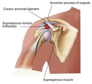
Shoulder impingement is a very common cause of shoulder pain, where a supraspinatus tendon inside the shoulder rubs or catches on nearby tissue and/or bone(acromium) as you lift your arm. Your shoulder is made up of several joints, muscles and tendons which allow the great range of motion in your arm. There are three bones in the shoulder joint: your upper arm bone, your shoulder blade, and your collarbone . Your arm is kept in your shoulder socket by your rotator cuff muscles, along with ligaments and the joint capsule which envelopes the shoulder. There is a lubricating sac called a bursa between the supraspinatus muscle of the rotator cuff where the supraspinatus travels under the bone on top of your shoulder (acromion). The bursa allows the supraspinatus tendon to glide freely under the acromium when you move your arm. When the bursa becomes swollen and inflamed, the condition is called bursitis and is one cause of shoulder impingement. Bone spurs within the shoulder joint can also contribute to impingement, as can forward curving shoulders due to years of bad posture. Sometimes due to impingement and/or bone spurs the supraspinatus muscle or tendon can tear. It can also happen during an accident like a shoulder dislocation or falling on a shoulder etc. Those who do repetitive lifting or overhead activities using the arm, such as paper hanging, construction, or painting are also susceptible.


Symptoms of shoulder impingement

You may have pain and stiffness when you lift your arm. There may also be pain when the arm is lowered from an elevated position. Early symptoms may be mild. Patients frequently do not seek treatment at an early stage. These symptoms may include:

  • Minor pain that is present both with activity and at rest
  • Pain radiating from the front of the shoulder to the side of the arm
  • Sudden pain with lifting and reaching movements
  • Athletes in overhead sports may have pain when throwing or serving a tennis ball

As the problem progresses, the symptoms increase:

  • Pain at night
  • Loss of strength and motion
  • Difficulty doing activities that place the arm behind the back, such as buttoning or zippering

If the pain comes on suddenly, the shoulder may be severely tender. All movement may be limited and painful.


Imaging Tests

Other tests which may help your doctor confirm your diagnosis include:

X-rays. Becauses x-rays do not show the soft tissues of your shoulder like the rotator cuff, plain x-rays of a shoulder with rotator cuff pain are usually normal or may show a small bone spur. A special x-ray view, called an “outlet view,” sometimes will show a small bone spur on the front edge of the acromion.

Magnetic resonance imaging (MRI) and ultrasound. These studies can create better images of soft tissues like the rotator cuff tendons. They can show fluid or inflammation in the bursa and rotator cuff. In some cases, partial tearing of the rotator cuff will be seen.

Treatment

The goal of treatment is to reduce pain and restore function. In planning your treatment, your doctor will consider your age, activity level, and general health.

TreatmentIn cases where there is a tear in the supraspinatus muscle or tendon, conservative treatment such as rest, physio etc rarely works. In milder cases rest along with rehab and correction of postural problems in the shoulder may work.Rest. Your doctor may suggest rest and activity modification, such as avoiding overhead activities.

Non-steroidal anti-inflammatory medicines. Drugs like ibuprofen and naproxen reduce pain and swelling in mild cases.

Physical therapy. A physical therapist will initially focus on restoring normal motion to your shoulder. Stretching exercises to improve range of motion are very helpful. If you have difficulty reaching behind your back, you may have developed tightness of the posterior capsule of the shoulder (capsule refers to the inner lining of the shoulder and posterior refers to the back of the shoulder). Specific stretching of the posterior capsule can be very effective in relieving pain in the shoulder.

Once your pain is improving, your therapist can start you on a strengthening program for the rotator cuff muscles.

Steroid injection.  Cortisone is a very effective anti-inflammatory medicine. Injecting it into the bursa beneath the acromion can relieve pain and reduce symptoms dramatically if the sole cause of the impingement is bursitis.


Surgical Treatment

When nonsurgical treatment does not relieve pain, your doctor may reoved. This is also known as a subacromial decompression. These procedures can be performed using either an arthroscopic or open technique.

Arthroscopic technique. In arthroscopy, thin surgical instruments are inserted into two or three small puncture wounds around your shoulder. Your doctor examines your shoulder through a fiberoptic scope connected to a television camera. He or she guides the small instruments using a video monitor, and removes bone and soft tissue. In most cases, the front edge of the acromion is removed along with some of the bursal tissue.

Your surgeon may also treat other conditions present in the shoulder at the time of surgery. These can include arthritis between the clavicle (collarbone) and the acromion (acromioclavicular arthritis), inflammation of the biceps tendon (biceps tendonitis), or a partial rotator cuff tear.

Open surgical technique. In open surgery, your doctor will make a small incision in the front of your shoulder. This allows your doctor to see the acromion and rotator cuff directly.

Rehabilitation. After surgery, your arm may be placed in a sling for a short period of time. This allows for early healing. As soon as your comfort allows, your doctor will remove the sling to begin exercise and use of the arm.


Physiotherapists in Tralee, Co. Kerry for all your physiotherapy needs. Phone 0867700191

Temperomandibular Joint Disorder
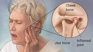
The temporomandibular joint works as a combination of hinge and sliding actions. It connects the jawbone to the skull on each side of the face.  The parts of the bones that interact in the joint are covered with cartilage and are separated by a small shock-absorbing disk, which normally keeps the movement smooth.  Temporomandibular  joint disorder (TMJD) can cause pain in your jaw joint and in the muscles that control jaw movement.

Symptoms

  • Pain in one or both of the temporomandibular joints
  • Difficulty or pain chewing
  • Locking of the joint, making it difficult to open or close your mouth
  • Clicking sound or grating sensation when you open your mouth or chew

Causes of  Temporomandibular Joint Disorder

Painful TMJ disorders can occur if:

  • The disk erodes or moves out of its proper alignment
  • The joint’s cartilage is damaged by arthritis
  • The joint is damaged by a blow or other impact

In many cases, however, the cause of TMJ disorders is unclear.

Treatment of  Temporomandibular Joint Disorder

In some cases, the symptoms of TMJ disorders may go away without treatment. Some of the following may also help.

Medications

  • Pain relievers and anti-inflammatories. These can help relieve pain and inflammation.
  • Muscle relaxants. These can help relax the jaw muscles.

Therapies

Nondrug therapies for TMJ disorders include:

  • Oral splints or mouth guards.  These devices worn at night while sleeping can help prevent grinding of the teeth.
  • Physical therapy.  Deep tissue massage work on neck and jaw muscles can be beneficial along with certain rehabilitation exercises.
  • Education. Education can help you understand the factors and behaviors that may aggravate your pain, so you can avoid them. Examples include teeth clenching or grinding, eating foods where you have to open the jaw wide like apples and burgers.

When conservative treatments fail, the following may be considered:

  • Arthrocentesis. This is a minimally invasive procedure that involves the insertion of small needles into the joint so that fluid can be irrigated through the joint to remove debris and inflammatory byproducts.
  • Injections. In some people, corticosteroid injections into the joint may be helpful. Infrequently, injecting botulinum toxin type A (Botox, others) into the jaw muscles used for chewing may relieve pain associated with TMJ disorders.
  • TMJ arthroscopy.  A small thin tube is placed into the joint space, an arthroscope is then inserted and small surgical instruments are used for surgery. TMJ arthroscopy has fewer risks and complications than open-joint surgery does, but it has some limitations as well.
  • Modified condylotomy. Modified condylotomy addresses the TMJ indirectly, with surgery on the mandible, but not in the joint itself. It may be helpful for treatment of pain and if locking is experienced.
  • Open-joint surgery. If your jaw pain does not resolve with more-conservative treatments and it appears to be caused by a structural problem in the joint, your doctor or dentist may suggest open-joint surgery (arthrotomy) to repair or replace the joint. However, open-joint surgery involves more risks than other procedures do.


For some handy self treatment tips, check out this video https://youtu.be/7b73yE0U2t0

Tmj manipulation video


Check out our website and feel free to contact us to discuss your condition or to set up an appointment.


Morton's Foot
Morton’s Foot

Morton’s foot and pyridoxal 5′-phosphate deficiency: genetically linked traits.

I came across an extremely interesting article lately regarding Morton’s foot and vitamin B6 deficiency. A lot of what I say in the article comes from scientifically published research referenced at the end of the article. Apologies if some of it is a bit technical. I have tried to simplify it while keeping the content intact.

What is Morton’s foot?

A Morton’s foot  also  called Morton’s toe, is a condition characterized by a longer second toe.

Pathophysiology

Morton’s Toe will cause an individual to have abnormal or over pronation.  It is this pronation that is the ultimate cause or contributing factor to most of the problems not only of the foot but also of the whole body.

Normal Pronation is a series of motions the foot must have, so that it can absorb the shock of meeting the ground. It must be able to do this, in order to adapt and adjust to the new walking surfaces it has just met. This adjustment should only last a fraction of a second to allow the foot to slow down; absorb the shock of your body weight in order to adjust and adapt to the walking surface. If this adjustments last longer, the foot will then begin to abnormally pronate and to correct itself. This is the start of a “chain reaction” that puts the foot under a lot of abnormal stress and strain, causing  Bunions, Heel Spurs, plantar fasciitis, Corns, Callouses, ingrown toenails and numerous other foot problems.


Vitamin B6 deficiency

Vitamin B6 is an essential vitamin needed for many chemical reactions in the human body. It exists as several active forms but pyridoxal 5′-phosphate (PLP) is the phosphorylated form needed for transamination, deamination, and decarboxylation. PLP is important in the production of neurotransmitters, acts as a Schiff base and is essential in the metabolism of homocysteine, a toxic amino acid involved in cardiovascular disease, stroke, thrombotic and Alzheimer’s disease. Nichols and Gaiteri(2014) showed the connection between a deficit of pyridoxal 5′-phosphate and the physical foot deformity known as the Morton’s foot. Morton’s foot has been associated with fibromyalgia/myofascial pain syndrome. PLP deficiency also plays a role in impaired glucose tolerance and may play a much bigger role in the obesity, diabetes, fatty liver and metabolic syndrome. Without the Schiff-base of PLP acting as an electron sink, storing electrons and dispensing them in the mitochondria, free radical damage occurs.


Summary

To put this all very simply : Vitamin B6 is an essential vitamin needed for many chemical reactions that take place within the human body. This vitamin is obtained from your diet. The genetically linked condition of ”Morton’s toe” has been linked to the inability to convert Vitamin B6 into the active form pyridoxal 5′-phosphate needed for cetain chemical processes in the human body. This in turn can lead to conditions seen with vitamin B6 deficiency. Some of these conditions include:

  • Anemia
  • Skin/Hair/Nail problems
  • Depression/Anxiety
  • Nerve damage
  • Pain syndrome development
  • Systemic Inflammation
  • Circulation problems, including oxygen transport
  • Hormone Issues
  • Seizures

Do not take a B6 supplement, or “b-vitamin complex” and expect it to help. It won’t.
You need the activated form of B6 called P5P. This is the only form that your body can use.


Reference:

Supplementation with PLP, L5-MTHF, B12 and trimethylglycine should be used in those patients with hyperhomocysteinemia and/or MTHFR gene mutation.(Trent W. Nichols, Christopher Gaiteri ,Published in Medical hypotheses 2014, DOI:10.1016/j.mehy.2014.09.003).


Physiotherapists in Tralee.  Open early until late. Phone 086-7700191